
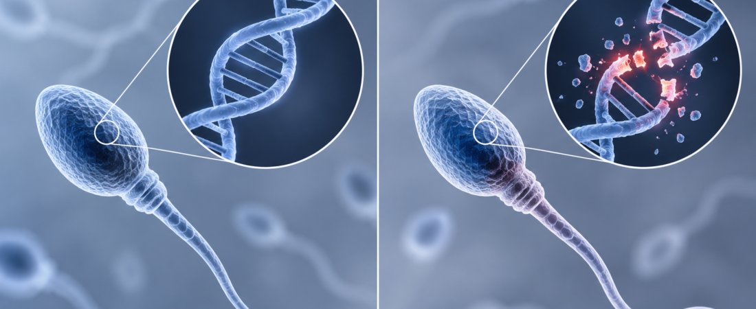
Blastocyst chromosome-abnormality rates were 38.19% in the high sperm DNA fragmentation group and 24.42% in the low group. Among couples whose female partner was at least 35, the corresponding rates were 52.02% and 38.75%. The researchers identified an association, not proof of causation.
Men undergoing fertility investigations may be told that their sperm DNA fragmentation index (DFI) is high. This can be confusing when sperm count, motility and appearance otherwise seem acceptable.
If sperm DNA is damaged, does that also affect the chromosomes of embryos created through IVF?
A large study examining assisted reproduction records over an eight-year period offers relevant evidence. Blastocysts created with sperm from men with a high DFI had more numerical chromosome abnormalities than those from men with a low DFI. The association was more pronounced when the female partner was at least 35 or when semen quality was severely impaired.
DNA damage is not fully captured by count and motility
Sperm DNA fragmentation refers to damage or breaks in DNA strands inside the sperm head.
A routine semen analysis primarily examines sperm numbers, movement and the proportion with normal morphology. A sperm that moves well and looks normal does not necessarily contain completely intact DNA.
One measure used to assess this is the DNA fragmentation index. In simple terms, it quantifies the proportion of sperm showing evidence of DNA damage in the tested sample.
Whether a high DFI is associated with the chromosome status of embryos has been less clear. Previous studies have produced conflicting findings. Some reported an association and others did not, while small samples and differences in testing methods have made firm conclusions difficult.
A total of 5,637 couples were screened; 3,948 were analyzed
The researchers screened 5,637 couples who received assisted reproductive treatment at a large reproductive medicine center between January 2016 and March 2023. Of these, 3,948 met the study criteria and had the necessary complete data for analysis.
Sperm DNA damage was measured using the Sperm Chromatin Structure Assay (SCSA). The researchers examined the relationship between DFI and blastocyst chromosome abnormalities, including analyses according to the partners' ages, semen characteristics and treatment methods.
The observed abnormality rate was 24.42% in the low-DFI group and 38.19% in the high-DFI group. Put another way, roughly 24 of every 100 blastocysts in the low group had a chromosome abnormality, compared with about 38 in the high group.
The association remained after statistical adjustment for factors that could influence the results, including the partners' ages. The adjusted odds ratio for chromosome abnormalities in the high- versus low-DFI group was 1.35.
This should not be interpreted as a 35% increase in the probability of an abnormal embryo. An odds ratio does not directly compare probabilities. The observed rates of 24.42% and 38.19% offer a more straightforward description of what was found in the study groups.
The association was stronger when the female partner was at least 35
When the female partner was at least 35, chromosome abnormalities were found in 38.75% of blastocysts in the low-DFI group and 52.02% in the high group. That is approximately 39 versus 52 out of every 100 blastocysts.
The association persisted after adjustment for other factors, with an adjusted odds ratio of 1.61. Again, this does not mean that the probability was 61% higher.
Why might the female partner's age matter alongside sperm DNA damage?
After fertilization, some damage to DNA contributed by sperm can be repaired through mechanisms involving the egg. The findings therefore raise the possibility that sperm DNA damage and characteristics of the egg interact during embryo development.
However, this study did not directly prove that eggs lose a particular repair ability after age 35 or that this mechanism causes sperm DNA damage to produce chromosome abnormalities. That explanation remains a biological hypothesis rather than a mechanism demonstrated by this analysis.
Men with severely impaired semen quality also showed a stronger association
Differences were also seen according to semen characteristics.
Among men with severe oligoasthenoteratozoospermia (SOAT), which combines a very low sperm count with severely reduced motility and a low proportion of normally shaped sperm, blastocyst chromosome-abnormality rates were 18.03% in the low-DFI group and 36.81% in the high group.
The adjusted odds ratio was 1.99. The relationship between high DFI and embryo chromosome abnormalities was therefore more pronounced in this group with severely abnormal conventional semen parameters.
Among men younger than 40, high DFI was also associated with a lower likelihood of producing good-quality blastocysts.
A high DFI does not mean normal embryos are impossible
The findings must not be read as showing that high DFI inevitably produces abnormal embryos, or that a man with a high DFI cannot contribute to a chromosomally normal embryo.
First, this was a single-center retrospective cohort study using past treatment records, not a randomized trial. The researchers adjusted statistically for several factors, including the partners' ages and semen characteristics, but acknowledged that unmeasured influences could not be completely excluded.
Second, the group with a moderate DFI did not have a clearly different chromosome-abnormality rate from the low-DFI group. The study therefore does not establish that every small increase in DFI produces a proportional increase in embryo abnormalities.
Third, the findings show an association. They do not demonstrate that high sperm DNA fragmentation directly caused the chromosome abnormalities.
The study is nevertheless informative because it examined sperm DNA quality, which is not fully described by count, motility and morphology, in a relatively large cohort and related it to embryo chromosome findings.
When embryo chromosome abnormalities are discussed in IVF, maternal age and egg quality often come first. This study suggests that the extent of sperm DNA damage may also be associated with embryo chromosome status.
That relationship was more evident when the female partner was at least 35 or semen quality was severely impaired. The findings support examining possible contributions from both eggs and sperm, while retaining the limits of observational evidence.
This article is based on research by investigators from Central South University and the Reproductive & Genetic Hospital of CITIC-Xiangya, China, and collaborating institutions, published in Human Reproduction Open on September 16, 2026. The study screened 5,637 couples and included 3,948 in the final analysis.
This article explains recent research for general readers. It does not replace medical judgment in interpreting an individual's test results or deciding whether a particular fertility test or treatment is appropriate.
Research references
High sperm DNA fragmentation is associated with increased embryonic chromosomal abnormality: a retrospective study of 8 years involving 5637 couples undergoing ART. Human Reproduction Open, September 16, 2026. Accepted manuscript. DOI: 10.1093/hropen/hoag082.
Related reading
The images used in this article were created using generative AI (ChatGPT, OpenAI). Any people depicted are fictional, not real individuals.
Copyright © 2026 Fertility Information TokTok Fertility News | THE FERTILITY NOTES. All rights reserved. Unauthorized republication, reproduction, redistribution, scraping, AI training, or use of any content, including articles, photographs, and images, as data is strictly prohibited. Violations may result in legal liability.