
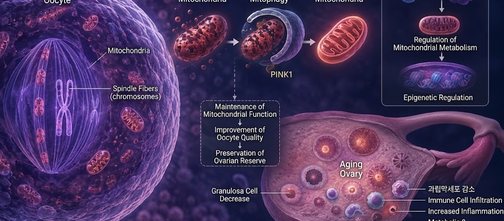
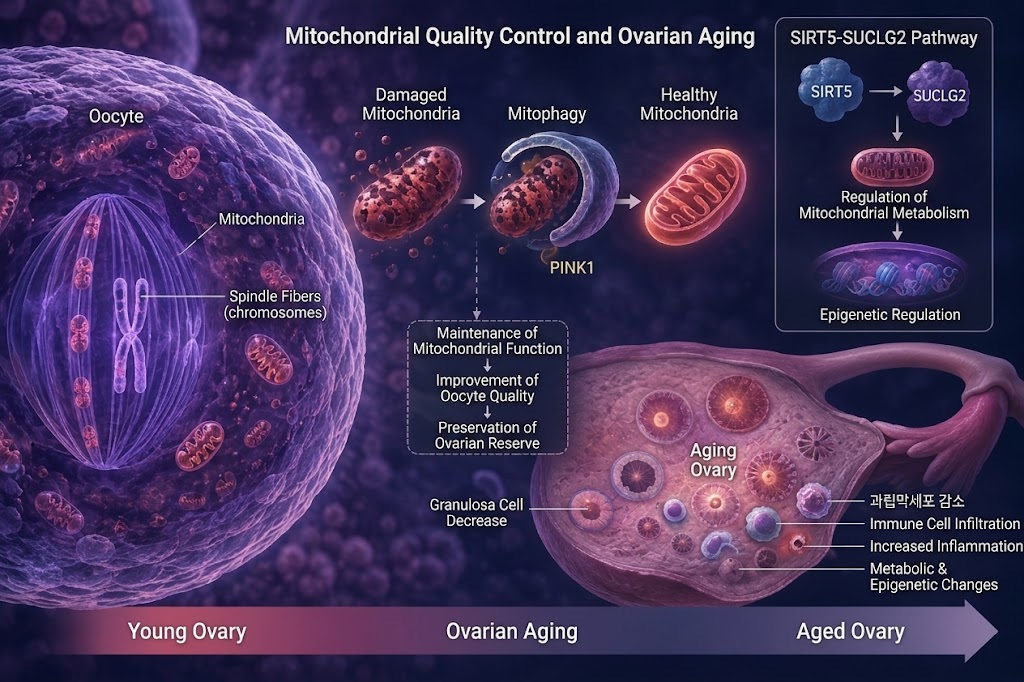
- PINK1-Driven Mitophagy Deficiency Causes Ovarian Reserve and Oocyte Competence to Fall: Loss of mitochondrial clearance increases cytoplasmic fragmentation and disrupts meiotic spindle assembly
- SIRT5-SUCLG2 Pathway Bridges Mitochondrial Bioenergetics and Nuclear Epigenetics: Mitochondrial TCA cycle dysfunction alters nuclear acetyl-CoA pools and histone acetylation profiles
- Reframing Ovarian Aging: Expanding from numerical “follicle depletion” to an integrated model of bioenergetic, inflammatory, and epigenetic tissue senescence
The human ovary exhibits an accelerated biological aging rate compared to other somatic organs. The traditional paradigm has long explained this through the finite exhaustion of primordial follicles until menopause.
Recent research, however, reveals that ovarian aging is driven not merely by the passive depletion of oocyte inventory, but by the progressive breakdown of the internal mitochondrial quality control systems that maintain the cellular power plants of oocytes and their supporting somatic cells.
Two 2026 studies place mitochondria at the center of reproductive senescence: one investigating mitophagy (the selective autophagy and degradation of dysfunctional mitochondria), and another showing how mitochondrial metabolic shifts directly reshape nuclear gene expression through epigenetic remodeling. Together, they demonstrate that ovarian aging is an interconnected, organ-level cascade spanning mitochondrial bioenergetics, chronic inflammation, and epigenetics.
Failure of the Clearance System: The PINK1 Pathway
Mitochondria generate the cellular ATP required for gamete maturation. However, damaged or depolarized mitochondria release excessive reactive oxygen species (ROS) and pro-apoptotic factors. To maintain cellular homeostasis, cells identify and degrade dysfunctional mitochondria via selective autophagy (mitophagy).
A study published in Free Radical Biology and Medicine (May 2026 issue; online February 2026) investigated the role of PINK1—a serine/threonine kinase that senses mitochondrial damage—in ovarian aging:
- Phenotypic Collapse in PINK1-Knockout Models: Mice lacking PINK1 exhibited significantly reduced ovarian weight, accelerated ovarian reserve depletion, increased granulosa cell apoptosis, and lower ovulation efficiency.
- Degraded Oocyte Competence: Oocytes from PINK1-deficient ovaries showed abnormal mitochondrial distribution, reduced membrane potential, elevated cytoplasmic fragmentation, and severe meiotic spindle assembly defects, leading to aneuploidy and early embryonic developmental arrest.
- Non-Canonical PINK1 Signaling: Notably, knockout of Parkin—the classic downstream E3 ubiquitin ligase in somatic mitophagy—did not fully replicate the severity of PINK1 deficiency. This suggests PINK1 regulates ovarian mitochondrial quality control via unique, non-canonical pathways distinct from other tissues.
Energy Infrastructure Over Simple Oocyte Count
These findings indicate that ovarian reserve cannot be evaluated solely as a numerical count of remaining follicles.
To complete meiotic division, align chromosomes accurately on the metaphase plate, fertilize, and sustain early embryonic cleavage before genomic activation, the oocyte relies entirely on its maternal mitochondrial pool. When mitophagy fails to clear damaged mitochondria, the bioenergetic capacity of the oocyte declines even while the physical follicle remains present in the ovary.
Mitochondrial-to-Nuclear Epigenetic Crosstalk: SIRT5-SUCLG2
A complementary study published in Nature Communications (July 15, 2026) utilized single-cell transcriptomics on aging ovaries to map the interplay between mitochondrial metabolism, cellular senescence, and histone modifications.
The researchers identified the SIRT5-SUCLG2 axis as a key metabolic-epigenetic regulator:
- SIRT5 Regulatory Function: SIRT5 (a desuccinylase/demalonylase) maintains the stability and enzymatic activity of SUCLG2 (succinate-CoA ligase GDP-forming subunit beta), an enzyme in the mitochondrial TCA cycle.
- Metabolic Reprogramming: Loss or downregulation of SIRT5 disrupts TCA cycle flux and alters intracellular concentrations of key metabolic intermediates, particularly acetyl-CoA.
- Epigenetic Disruption: This mitochondrial disruption depletes the nuclear acetyl-CoA pool, leading to widespread aberrant histone acetylation and altered transcription of genes regulating cell survival and senescence.
This confirms a direct functional bridge where mitochondrial metabolic dysfunction directly reprograms nuclear gene expression.
The Emerging Paradigm: Ovarian Aging as a Multi-Tissue Network
Connecting these findings highlights how reproductive endocrinology is shifting its conceptual framework of ovarian aging:
| Traditional Paradigm | Emerging Metabolic-Epigenetic Paradigm |
| Primary Driver | Passive depletion of oocyte pool (“inventory exhaustion”) |
| Mechanistic Focus | Endocrine decline (AMH, Inhibin B, Estradiol) |
| Nuclear vs. Cytoplasm | Meiotic cohesin deterioration & chromosomal aneuploidy |
| Cellular Target | Oocyte alone |
Ovarian aging is an active biological process where compromised mitochondrial clearance (loss of mitophagy), metabolic reprogramming, chronic low-grade inflammation (inflammaging), and downstream epigenetic deregulation interact to degrade the follicular environment.
Study Limitations and Translational Horizons
While these molecular insights are significant, essential clinical boundaries apply:
- Preclinical Evidence: Both the PINK1 mitophagy study and the SIRT5-SUCLG2 pathway analyses were conducted in animal models and in vitro cellular systems.
- No Proven Reversal Therapies: Current data does not yet prove that administering specific mitophagy enhancers or mitochondrial-targeted supplements will restore human ovarian reserve or delay the onset of natural menopause.
Nevertheless, understanding these pathways provides actionable targets for future reproductive medicine. Rather than viewing reproductive aging as an unalterable biological clock, elucidating mitochondrial clearance and epigenetic remodeling identifies distinct molecular mechanisms that may one day be targeted to maintain oocyte competence and follicular health.
Medical Sources & Literature References
- Journal: Free Radical Biology and Medicine (May 2026; Online February 2026) — PINK1 deficiency impairs mitochondrial quality control and accelerates ovarian aging
- Journal: Nature Communications (Published July 15, 2026) — The SIRT5-SUCLG2 axis couples mitochondrial metabolic dysfunction to epigenetic histone modifications during ovarian aging
※ This article was synthesized based on peer-reviewed studies published in Free Radical Biology and Medicine and Nature Communications (2026) alongside literature in cellular bioenergetics and reproductive biology. It does not replace individualized clinical diagnosis or medical care, and specific treatment decisions should always be made in consultation with a qualified reproductive specialist.
※ The images associated with this article were generated using generative AI (ChatGPT, OpenAI) as illustrative visual references and do not depict real individuals.