

Estimating Chromosomal Status from a Subset of Blastocyst Cells… Not an Evaluation of the Entire Embryo Case Reports of Mosaicism Detected in Conception Products Following ‘Normal’ PGT-A Diagnosis Small Genetic Variants, Uterine, and Placental Factors Remain Beyond PGT-A’s Reach
“It is a PGT-A euploid embryo.”
For patients undergoing in vitro fertilization (IVF), few findings bring greater relief. After harvesting and fertilizing eggs, culturing them to the blastocyst stage, and passing preimplantation genetic testing for aneuploidy (PGT-A), it is easy to feel as though the most critical hurdle has been cleared.
Yet, miscarriages still occur even after transferring a euploid embryo and confirming pregnancy. Why does this happen when the chromosomes were reported as normal?
What ‘PGT-A Normal’ Truly Means
The answer lies in the clinical definition of a “PGT-A normal” embryo.
A PGT-A euploid embryo does not mean an embryo that is genetically flawless across every biological metric. Rather, it means that no major numerical chromosomal abnormalities were detected within the biopsied cells under current testing methodologies and laboratory reporting thresholds.
PGT-A does not analyze the entire embryo. Typically, DNA is extracted from a small sample of cells biopsied from the outer trophectoderm (TE) of a Day 5 or Day 6 blastocyst. These cells primarily go on to form the placenta. The test does not examine every individual cell, nor does it sample the inner cell mass (ICM) that develops into the fetus.
This biological reality creates room for mosaicism. A single embryo can harbor a mixture of chromosomally normal and abnormal cell lines, meaning a biopsy of a few trophectoderm cells may not perfectly represent the chromosomal constitution of the whole embryo.
The American Society for Reproductive Medicine (ASRM) notes that mosaicism reporting criteria can vary based on next-generation sequencing (NGS) platforms, bioinformatics software, and individual laboratory cutoffs. Intermediate abnormal signals can reflect true biological mosaicism or technical noise inherent to the testing platform.
Indeed, a 2026 study published in the international journal Frontiers in Reproductive Health reported cases of two women who each underwent single euploid embryo transfers, experienced first-trimester miscarriages, and subsequent chromosomal testing of the products of conception revealed mosaic chromosomal abnormalities.
In one case, initial PGT-A testing displayed a roughly 10% abnormal signal on chromosome 8, which was categorized as “euploid/normal” according to that laboratory’s diagnostic threshold. Following the miscarriage, genetic analysis of the tissue identified chromosome 8 mosaicism at a substantially higher percentage.
Two case reports cannot explain every instance of euploid pregnancy loss. However, they clearly illustrate that a “normal” PGT-A result does not serve as an absolute, whole-embryo biological guarantee.
Genetic Lesions Beyond Aneuploidy
PGT-A is not designed to detect all genetic abnormalities.
Its primary clinical objective is screening for aneuploidy—the gain or loss of whole chromosomes. While modern NGS platforms can identify segmental deletions/duplications above certain size thresholds and intermediate mosaic signals, they do not detect single-gene mutations, epigenetic errors, or subtle microdeletions and microduplications.
A 2025 whole-genome sequencing (WGS) study published in Nature identified a higher burden of pathogenic, small-scale de novo genetic variants in early pregnancy loss specimens compared to healthy adult controls. Another meta-analysis detected pathogenic copy number variations (CNVs) in a subset of spontaneous miscarriages presenting with normal chromosome counts.
While these studies did not exclusively analyze PGT-A-tested euploid transfers, they underscore that a normal chromosome count alone cannot rule out all genetic drivers of miscarriage.
Embryo Viability, Uterine, and Placental Variables
Even among euploid embryos, developmental potential varies. Euploid embryos can differ significantly in morphokinetics, the day they reach full blastocyst expansion, and the morphological grading of the inner cell mass and trophectoderm.
Beyond the embryo, the uterus and maternal-fetal interface introduce another major set of variables. Following implantation, an embryo must interact with the endometrium, remodel maternal spiral arteries, and establish a functional placenta. Uterine structural anomalies, thrombophilias, endocrine and metabolic disorders, and maternal immunological factors can all disrupt ongoing pregnancy maintenance.
In clinical practice, the risk of pregnancy loss persists after transferring euploid embryos. A 2023 study analyzing 8,672 implanted euploid embryos reported a spontaneous miscarriage rate of 8.9%. While this rate is significantly lower than that observed with mosaic or unbiopsied embryos, it is not zero.
Clinical Takeaways and Perspective
These limitations do not diminish the clinical utility of PGT-A. It remains an important tool for filtering out whole-chromosome aneuploidies—the leading cause of early pregnancy loss—and prioritizing embryos for transfer.
However, viewing PGT-A as a test that “guarantees an embryo will never miscarry” creates an inevitable gap between expectations and clinical reality. The ASRM similarly highlights that routine application of PGT-A to all IVF patients does not universally improve cumulative live birth rates across every demographic.
When a miscarriage occurs following a euploid transfer, clinicians and patients should avoid immediately concluding that the PGT-A result was faulty, or conversely assuming that the embryo was flawless and the uterus was entirely at fault.
The outcome may stem from sampling discordance between the biopsy and the inner cell mass, genetic variants outside the diagnostic resolution of the assay, intrinsic metabolic competence, placental dysfunction, or maternal physiological factors. In many cases, early losses remain clinically unexplained.
Ultimately, a PGT-A euploid embryo is best understood as:
“An embryo for which major chromosomal aneuploidy risks have been substantially ruled out within the limits of current testing,” rather than “an embryo immune to miscarriage.”
By filtering out major chromosomal aneuploidies, PGT-A has brought reproductive medicine face-to-face with the questions that lie beneath: why certain chromosomally normal embryos still fail to sustain development, and how to better support the maternal-embryonic environment.
※ This article was synthesized based on clinical guidance from the American Society for Reproductive Medicine (ASRM), case reports of euploid miscarriage published in Frontiers in Reproductive Health (2026), whole-genome sequencing findings in pregnancy loss published in Nature (2025), and recent reproductive medicine literature. It does not replace individualized clinical diagnosis or treatment, and specific medical decisions should be made in consultation with a specialist physician.
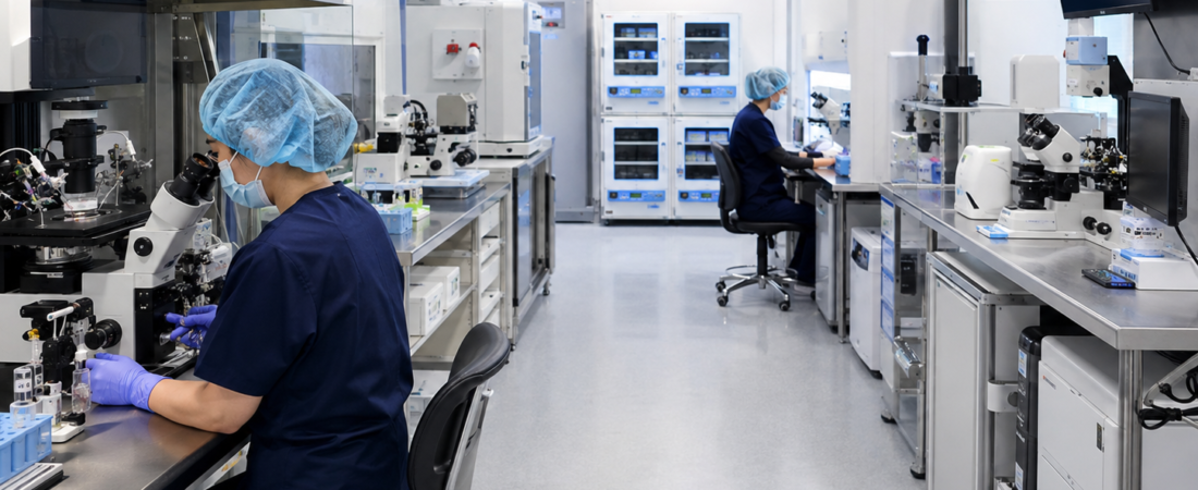
※ Images: Generated using generative AI (ChatGPT, OpenAI) as visual references and do not depict real individuals.