
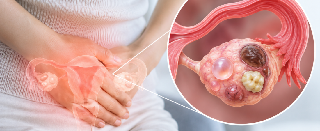
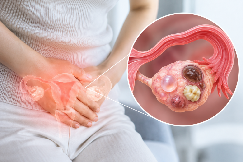
- A broad spectrum of pathology: From functional cysts that appear and resolve during normal ovulation to endometriomas, teratomas, and true benign or malignant neoplasms
- Different causes demand different treatments: Even under the shared label of an “ovarian cyst,” management diverges completely; women seeking fertility must approach surgery with heightened caution
- An ultrasound finding does not automatically equal cancer: Risk stratification relies on size, architecture, Doppler blood flow, and menopausal status
When a physician pauses during a pelvic ultrasound and mentions, “There is something visible on your ovary,” many women immediately fear the worst: cancer.
In reality, lesions discovered in the ovary span a far broader and more nuanced spectrum. Some are simple fluid-filled water cysts that emerge during this month’s ovulation only to resolve by the next; others are endometriomas filled with degraded, pooled blood, dermoid cysts containing fat and hair, benign neoplasms that grow independently of menstrual cycles, or, in rare instances, malignant tumors.
While they all appear as “an ovarian mass or cyst” on the surface, their underlying biological causes and clinical management could not be more different.
Appearing and Disappearing on Their Own: Functional Ovarian Cysts
The most common findings are functional ovarian cysts.
Every month, the ovaries recruit and mature follicles to release an egg. If a dominant follicle fails to rupture and release its oocyte, it can continue to expand, forming a follicular cyst. Conversely, if the corpus luteum—the temporary endocrine structure formed after ovulation—accumulates fluid or blood instead of regressing, it becomes a corpus luteum cyst.
The vast majority of functional cysts are entirely benign and typically regress spontaneously within a few weeks to several months without requiring medical intervention. This is precisely why, when a simple, smooth-walled water cyst is spotted on ultrasound, clinicians rarely rush to surgery, opting instead to re-evaluate the ovary after the patient’s next menstrual period.
Old Blood Pools Inside: Endometriomas (Chocolate Cysts)
For women planning a pregnancy, the ovarian endometrioma is of paramount clinical importance.
Commonly referred to as a “chocolate cyst,” an endometrioma forms when tissue biologically similar to the endometrial lining implants on the ovary. Subject to cyclical hormonal shifts, this ectopic tissue sheds and bleeds internally with each cycle. Because the blood has no exit route, it accumulates and degrades over months and years, taking on a thick, dark brown, syrup-like appearance reminiscent of melted chocolate.
An endometrioma is not merely a space-occupying lesion. It triggers persistent chronic inflammation and oxidative stress in the surrounding ovarian microenvironment, compromising local follicle health and contributing to subfertility.
However, surgically stripping the cyst capsule can inadvertently tear away normal ovarian cortex and damage functional primordial follicles, leading to a sharp post-operative drop in Anti-Mullerian Hormone (AMH). For women planning to conceive, deciding on surgery requires carefully balancing cyst size, pelvic pain, baseline ovarian reserve, and upcoming IVF plans.
Sudden Internal Bleeding: Hemorrhagic Cysts
Hemorrhagic cysts are another frequent finding. They occur when a fragile blood vessel ruptures within a physiological cyst—most often a corpus luteum—during or shortly after ovulation, filling the cavity with blood.
On an ultrasound monitor, the organizing clot and fibrin strands create an internal reticular pattern (often described as a “spiderweb” or “fishnet” appearance) that can look alarming to an untrained eye. In the majority of stable cases, however, the trapped blood is gradually broken down and reabsorbed by the body, allowing the cyst to resolve naturally over one to two cycles.
The primary risk arises if a hemorrhagic cyst ruptures and bleeds heavily into the peritoneal cavity. Significant intra-abdominal hemorrhage can trigger sudden, severe pelvic pain, dizziness, cold sweats, and peritoneal irritation, requiring prompt emergency evaluation.
Hair, Skin, and Fat: Mature Cystic Teratomas (Dermoid Cysts)
Perhaps the most startling lesion by name and content is the mature cystic teratoma, historically known as a dermoid cyst.
Arising from pluripotent germ cells responsible for generating gametes, these tumors have the biological capacity to differentiate into various germ layers. Consequently, their internal cavity can contain sebaceous fluid, adipose tissue, skin fragments, strands of hair, and occasionally calcified structures like bone or teeth. While this description sounds menacing, mature cystic teratomas are almost overwhelmingly benign.
The primary clinical complication stems from their physical weight and density. Because they are heavy and often mobile on an elongated infundibulopelvic ligament, they significantly elevate the risk of ovarian torsion—a condition where the ovary twists around its own blood supply. Ovarian torsion abruptly cuts off venous and arterial perfusion, presenting as sudden, excruciating unilateral lower abdominal pain, often accompanied by severe nausea and vomiting. Torsion constitutes an acute surgical emergency requiring prompt intervention to untwist the ovary and save viable tissue.
Looking Like Water Cysts but True Neoplasms: Serous and Mucinous Cystadenomas
Certain benign tumors grow independently of the ovulatory cycle. The most common varieties are serous cystadenomas and mucinous cystadenomas.
Serous cystadenomas are filled with thin, watery fluid, whereas mucinous cystadenomas contain thick, gelatinous mucus and have a propensity to grow to substantial sizes—sometimes spanning 15 to 20 centimeters or more. In their early stages, they produce virtually no symptoms, often going unnoticed until a patient presents with abdominal distension, pelvic fullness, or localized pressure on the bladder and bowel.
Unlike functional cysts, cystadenomas do not regress on their own. Depending on their diameter, structural complexity on imaging, patient age, and symptomatic burden, they are managed via periodic ultrasound surveillance or elective surgical excision.
An Abundance of Arrested Follicles: Polycystic Ovary Syndrome (PCOS)
Due to its terminology, Polycystic Ovary Syndrome (PCOS) is frequently confused with true ovarian cysts.
However, the multiple small, dark circles seen encircling the ovary on an ultrasound (the classic “string-of-pearls” sign) are not pathological cysts at all. They are small antral follicles whose maturation has temporarily arrested mid-cycle.
The root issue in PCOS is not a neoplasm, but an underlying neuroendocrine and metabolic imbalance marked by chronic anovulation, elevated luteinizing hormone, insulin resistance, and hyperandrogenism. PCOS must be recognized as an endocrine syndrome entirely distinct from solitary ovarian cystic lesions.
The Lesion Requiring the Greatest Vigilance: Ovarian Cancer
The most critical entity to rule out when evaluating an adnexal mass is a malignant neoplasm. However, it is vital to remember that an ultrasound notation of “an ovarian mass” is far from an immediate cancer diagnosis.
Sonographers and gynecologists systematically stratify malignancy risk using standardized imaging guidelines (such as the IOTA simple rules or O-RADS scoring). Key variables include:
- Patient Context: Menopausal status and age (the baseline probability of malignancy increases significantly post-menopause).
- Internal Architecture: Presence of solid tissue components, thick internal septations (> 3 mm), or papillary projections growing into the cyst cavity.
- Vascularity: Color Doppler evaluation showing low-resistance, high-velocity blood flow within solid nodules.
- Secondary Findings: Free peritoneal fluid (ascites) or peritoneal nodularity.
When imaging reveals suspicious complex features, clinicians promptly expand testing to include serum tumor markers (such as CA-125, HE4, and the ROMA algorithm) alongside pelvic contrast-enhanced MRI or CT imaging.
Modern gynecologic oncology has also revolutionized our understanding of where these tumors originate. A large body of molecular evidence confirms that the vast majority of High-Grade Serous Ovarian Carcinomas (HGSOC)—the most lethal and common form of ovarian cancer—do not originate in the ovary itself, but arise from precursor lesions in the fimbriated end of the fallopian tubes (STIC, or Serous Tubal Intraepithelial Carcinoma). This discovery has fundamentally shifted cancer prevention strategies, paving the way for opportunistic salpingectomy (removing the fallopian tubes while preserving healthy ovaries) during routine pelvic surgeries.
Beyond the Ultrasound Screen
Hearing that you have “a cyst on the ovary” confirms nothing on its own.
The first step is determining the biological nature of the mass: is it clear fluid, trapped blood, ectopic endometrium, sebaceous tissue, or a proliferative neoplasm? The ovary is an intensely dynamic organ that grows, ruptures, and repairs tissue every single month. A lesion that was absent yesterday can form today, and a cyst seen today may disappear completely after the next menstrual cycle. What matters far more than the word “cyst” on a report is understanding the exact nature of the lesion and observing how it behaves over time.
For women planning a pregnancy or undergoing assisted reproduction, surgical decisions should never be based solely on raw centimeter measurements. Two cysts of identical diameter demand entirely different management based on their histological subtype, proximity to functional cortex, the patient’s baseline ovarian reserve, and upcoming egg retrieval plans.
The clinical objective is never simply to “make the mass vanish from the ultrasound image.” For a woman who still hopes to carry a pregnancy, viable follicles must remain once the lesion is addressed.
The defining question should never be “How cleanly can we strip this cyst away?” but rather “How much of my healthy ovary can we preserve while treating it?”
※ This article was synthesized based on clinical practice bulletins from the American College of Obstetricians and Gynecologists (ACOG) on the “Evaluation and Management of Adnexal Masses” alongside the European Society of Human Reproduction and Embryology (ESHRE) Endometriosis Guidelines published in Human Reproduction Open. It does not replace individualized clinical diagnosis or medical care, and specific treatment decisions should always be made in consultation with a qualified gynecologist or reproductive endocrinologist.
※ The images associated with this article were generated using generative AI (ChatGPT, OpenAI) as illustrative visual references and do not depict real individuals.