
- 3D Quantitative Mapping of 85,000+ Oocytes Across Intact Cleared Ovaries: First whole-organ spatial capture of reproductive aging across the lifespan
- Activation Fraction Remains Constant Despite Severe Pool Depletion: The proportion of dormant oocytes entering the growing pool remains steady, pointing to organ-level feedback mechanisms
- Spatial Niches and Density Dictate Activation: Ovarian reserve redefined beyond a simple “static egg count” to an actively regulated spatial ecosystem
Does the female ovary simply lose stored eggs through passive, random attrition as age advances?
A whole-organ imaging study has revealed a fundamentally different biological model. Even as the total oocyte pool declined steeply with reproductive age, the proportion of dormant oocytes transitioning into the active growth phase remained remarkably constant. This suggests that ovarian aging is not mere unorganized exhaustion, but an actively regulated organ-level process where the ovary monitors its reserve and orchestrates follicle activation.
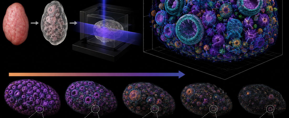
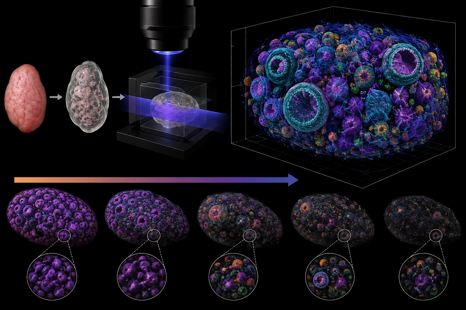
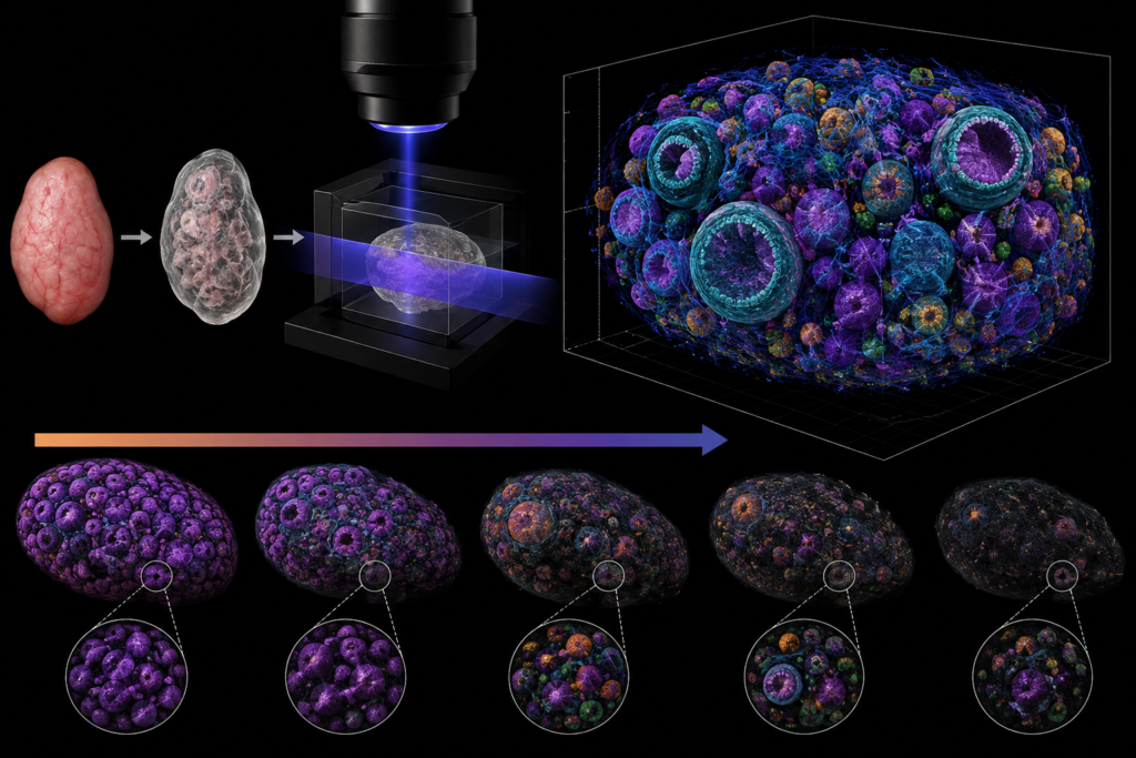
In a study published on August 12, 2026, in the international journal Nature Aging, an international research team led by the Centre for Genomic Regulation (CRG) in Spain utilized tissue optical clearing, light-sheet fluorescence microscopy, and artificial intelligence to construct a 3D spatial map tracking the exact coordinates, volumes, and developmental stages of over 85,000 individual oocytes across the lifespan of mice.
Unlike traditional two-dimensional histological sectioning—which captures only fragmented, physical slices—this approach analyzed the entire intact ovary as a continuous three-dimensional architecture.
A Steady Activation Rate Despite Depleted Reserves
The researchers’ most striking finding centered on how the ovarian reserve is mobilized over time.
While the absolute count of primordial follicles dropped dramatically with advancing age, the fraction of dormant oocytes recruited into the growing pool remained consistent throughout reproductive life.
Historically, ovarian aging has been conceptualized as passive, steady depletion—a timer simply running down until the follicle reservoir is empty. Clinically, ovarian reserve is estimated via surrogate markers such as Anti-Müllerian Hormone (AMH) and Antral Follicle Count (AFC), which quantify available pool size.
However, these 3D findings indicate that the ovary maintains an intrinsic, organ-level regulatory balance that scales activation rates to the total available reserve rather than letting follicles activate at random.
Spatial Distribution and Follicular “Density Niches”
Oocytes were not randomly scattered throughout the ovarian cortex:
- Spatial Clustering Along Structural Axes: Follicles exhibited non-random spatial orientation along defined anatomical axes.
- Density-Driven Activation: Regions with a high local density of primordial follicles displayed significantly higher rates of recently activated oocytes.
This demonstrates that primordial follicle emergence from dormancy is not governed solely by isolated cell-intrinsic signals. Instead, local follicular packing density, mechanical tissue cues, and microenvironmental niches within the ovarian architecture exert significant influence on whether a follicle remains dormant or initiates folliculogenesis.
Additionally, whole-organ volumetric profiling revealed a distinct bimodal size distribution:
- Beyond the large population of small, quiescent primordial oocytes, a separate distinct cluster emerged around $60\ \mu\text{m}$ in diameter.
- This indicates the presence of conserved developmental bottlenecks or temporary kinetic pauses during early follicular growth, a pattern that persisted even in aged ovaries with severely reduced reserves.
Shifting Paradigms: From “How Many Remain?” to “How Are They Regulated?”
These findings reframe a fundamental biological question in reproductive endocrinology: How does the ovary select a minute fraction of primordial follicles for growth while keeping the vast majority quiescent for decades?
Rather than individual follicles making autonomous activation decisions, the data supports the existence of an overarching, organ-level coordination system that integrates total pool size, local spatial density, and microenvironmental signals.
This perspective opens new avenues for investigating ovarian pathologies:
- Premature Ovarian Insufficiency (POI) & Early Menopause: Investigating whether rapid reserve depletion stems from accelerated oocyte death or a breakdown in the spatial brakes governing follicle activation.
- Fertility Preservation: Understanding the spatial niche factors required to sustain follicle dormancy or induce coordinated in vitro activation (IVA).
Study Limitations and Translational Horizon
- Murine Model: The primary longitudinal quantitative maps were generated in rodents.
- Human Validation: While the researchers confirmed that this 3D optical clearing and automated quantification pipeline can be applied to human ovarian cortical tissue, longitudinal validation across the entire human reproductive lifespan is still required.
- Clinical Boundary: These findings represent fundamental cell biology and spatial physiology; they do not yet translate into immediate clinical tools for predicting the exact age of menopause or altering ovarian stimulation protocols.
Conclusion
The biological clock of the ovary does not simply tick down by counting lost numbers. Ovarian reserve is not merely a static bucket of eggs; it is a dynamically regulated, spatially organized organ ecosystem.
By capturing the spatial order hidden beyond 2D histological sections, this 3D map demonstrates that ovarian aging follows defined organizational principles, challenging the conventional equation that “ovarian reserve equals simple egg count.”
Medical Source & Study Information
- Journal: Nature Aging (Published August 12, 2026)
- Study Title: Three-dimensional quantitative mapping of the ovary reveals spatial organization of oocyte activation during reproductive ageing
- Lead Institutions: Centre for Genomic Regulation (CRG), Spain, et al.
※ This article was synthesized based on the study published in Nature Aging (August 12, 2026) and literature in reproductive biology and ovarian aging. It does not replace individualized clinical diagnosis or medical advice, and specific medical decisions should always be made in consultation with a qualified reproductive specialist.
※ The images associated with this article were generated using generative AI (ChatGPT, OpenAI) as illustrative visual references and do not depict real individuals.