
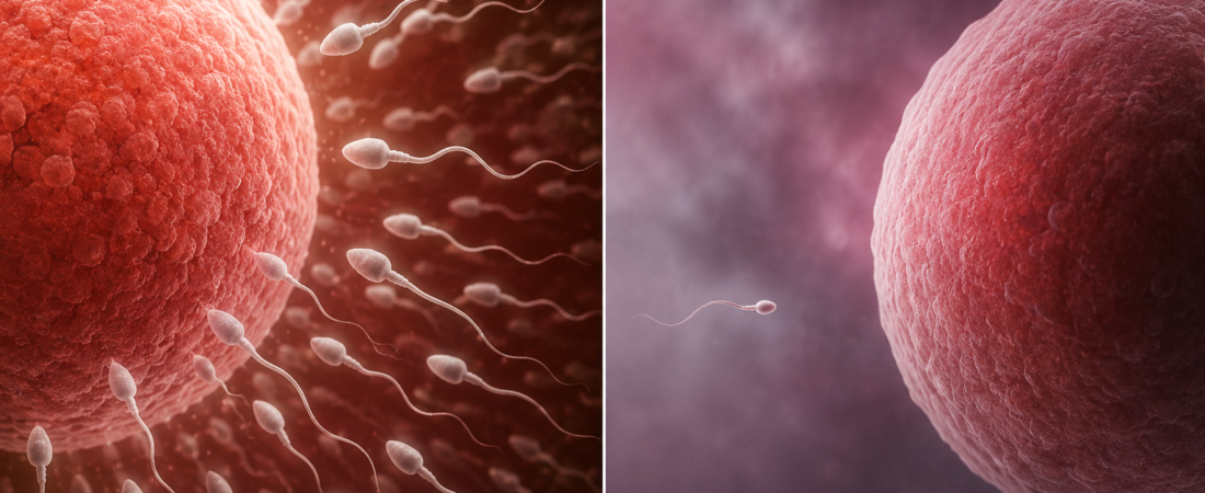
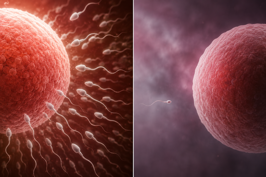
- The Underlying Mechanisms of Acquired Obstructive Azoospermia (OA)
- Testicular Spermatogenesis Remains Intact, But Blockages in the Epididymides, Vasa Deferentia, or Ejaculatory Ducts Result in Azoospermic Semen
- From Infections and Chronic Inflammation to Prior Vasectomies, Inguinal Surgeries, and Trauma… Identifying the Exact Obstructive Etiology Guides Treatment Strategy
When patients receive a diagnosis of azoospermia (the absence of measurable sperm in the ejaculate), the immediate assumption is often that the testes are failing to produce sperm. However, in many cases, testicular spermatogenesis functions normally, but the transport conduits are blocked along the pathway. This condition is clinically defined as Obstructive Azoospermia (OA).
In normal male reproductive physiology, spermatozoa are generated within the seminiferous tubules of the testes, transit to the epididymis for maturation and storage, travel through the vas deferens, and pass through the ejaculatory duct into the urethra, where they mix with secretions from the seminal vesicles and prostate gland during ejaculation. If a bilateral mechanical obstruction or disruption occurs anywhere along this tract, ejaculated semen will contain no sperm despite ongoing testicular production.
Elective vasectomy represents an intentional model of acquired obstructive azoospermia: transecting and ligating the vasa deferentia isolates sperm transport from the ejaculatory tract while testicular production persists.
Pathophysiology: A Problem of Transport, Not Production
Obstructive Azoospermia differs fundamentally from Non-Obstructive Azoospermia (NOA), which is characterized by intrinsic testicular failure or primary spermatogenic arrest:
- Endocrine & Physical Findings in OA: Testicular volume is typically normal (≥15–20 mL), and systemic gonadotropins—specifically Follicle-Stimulating Hormone (FSH)—and serum testosterone generally remain within normal reference ranges.
- Prognostic Advantage: Because spermatogenesis is intact, the clinical prognosis for achieving biological parenthood is substantially higher in OA than in NOA. Depending on the site and etiology of the blockage, options include microsurgical ductal reconstruction or surgical sperm retrieval combined with In Vitro Fertilization and Intracytoplasmic Sperm Injection (IVF-ICSI).
For men with previously documented fertility or normal semen parameters who subsequently develop azoospermia, acquired anatomical obstruction is the primary consideration.
Primary Causes of Acquired Obstructive Azoospermia
1. Genital Tract Infections and Inflammatory Fibrosis
Severe or recurrent infections of the male reproductive tract are a leading driver of acquired ductal obstruction:
- Epididymitis & Epididymo-orchitis: The epididymis consists of a single, microscopic, tightly convoluted tubule measuring approximately 6 meters in length. Acute or chronic inflammation can induce intraluminal scarring, mucosal adhesion, and complete fibrous occlusion of this delicate ductal system.
- Sexually Transmitted Infections (STIs): Pathogens such as Chlamydia trachomatis and Neisseria gonorrhoeae can ascend the genital tract, causing subclinical inflammation. Even after antibiotic clearance of the acute infection, residual post-inflammatory fibrosis and strictures may permanently occlude the epididymal lumen bilaterally.
2. Iatrogenic Surgical Injury
Prior surgical procedures in the groin, scrotum, or pelvis can inadvertently compromise the vas deferens:
- Elective Vasectomy: The most prevalent cause of intentional secondary OA.
- Inguinal Herniorrhaphy: Inguinal hernia repairs—especially pediatric hernia repairs, bilateral procedures, or adult repairs utilizing prosthetic polypropylene mesh—can result in vas deferens entrapment, mechanical transection, or chronic inflammatory fibrosis that obliterates the vasal lumen. Because childhood procedures may not be remembered clearly by the patient, pediatric surgical history is an essential diagnostic query.
- Scrotal & Pelvic Interventions: Hydrocelectomy, spermatocelectomy, or pelvic surgeries carry risks of incidental trauma to the epididymis or spermatic cord.
3. Physical Trauma
Severe blunt or penetrating trauma to the scrotum, perineum, or inguinal region (e.g., motor vehicle collisions, contact sports injuries, straddle trauma) can cause ductal laceration, scrotal hematomas, and secondary ischemic or cicatricial obstruction of the vasa deferentia or epididymides.
4. Ejaculatory Duct Obstruction (EDO)
Blockage at the distal end of the tract—where the vas deferens and seminal vesicle duct merge to form the ejaculatory duct within the prostate—can be caused by midline prostatic cysts (Müllerian or utricular cysts), ejaculatory duct calculi (stones), chronic prostatitis, or post-surgical scarring.
- Diagnostic Clues for EDO: Patients with complete EDO typically present with low-volume ejaculate (<1.0–1.5 mL), acidic semen pH (<7.2), and absent or low seminal fructose, as the alkaline, fructose-rich secretions of the seminal vesicles cannot discharge into the urethra.
Diagnostic Evaluation: Moving Beyond “No Sperm Found”
An azoospermia diagnosis should prompt a structured investigative workup to differentiate OA from NOA and localize the anatomical level of obstruction:
- Centrifuged Semen Analysis: Confirming azoospermia requires high-speed centrifugation (3,000×g for 15 minutes) and microscopic pellet examination to rule out severe cryptozoospermia.
- Physical Examination: Palpation of bilateral testicular consistency and size, presence and turgidity of the epididymides (a dilated, “indurated” epididymis suggests distal obstruction), and continuous palpation of the vasa deferentia (ruling out Congenital Bilateral Absence of the Vas Deferens [CBAVD]).
- Endocrine Profiling: Measuring serum FSH, LH, and Total Testosterone. Normal FSH in the presence of azoospermia and normal-sized testes strongly points toward an obstructive etiology.
- Imaging Modalities:
- Scrotal Doppler Ultrasound: Assesses testicular parenchymal architecture, epididymal tubular dilation, and associated varicoceles or spermatoceles.
- Transrectal Ultrasound (TRUS): Indicated when EDO is suspected; visualizes seminal vesicle dilation (>15 mm anteroposterior diameter), midline prostatic cysts, or intraductal calcifications.
Clinical Management: Reconstruction vs. Sperm Retrieval with ICSI
Once acquired OA is diagnosed, therapeutic management follows two primary pathways:
- Microsurgical Ductal Reconstruction:
- Vasovasostomy (VV) or Vasoepididymostomy (VE): High-magnification microsurgical re-anastomosis (e.g., following vasectomy or localized epididymal blockages) can restore patency and allow natural conception.
- Transurethral Resection of the Ejaculatory Ducts (TURED): Endoscopic resection of obstructing cysts or calcifications in confirmed EDO cases.
- Surgical Sperm Retrieval + IVF-ICSI:
- When surgical reconstruction is technically unfeasible, anatomically unfavorable, or when the female partner has advanced maternal age or diminished ovarian reserve requiring rapid ART intervention.
- Techniques include Percutaneous/Microsurgical Epididymal Sperm Aspiration (PESA/MESA) or Testicular Sperm Aspiration/Extraction (TESA/TESE). Because spermatogenesis is normal, sperm retrieval success rates in OA approach nearly 100%.
Summary
Acquired Obstructive Azoospermia represents a condition where sperm production is preserved, but the outflow tract has been mechanically interrupted.
For men who develop azoospermia later in life or after previously confirmed fertility, evaluating the integrity of the reproductive conduits—accounting for remote infections, surgeries, and trauma—is essential for selecting between microsurgical reconstruction and assisted reproduction.
Medical Guidelines & Clinical References
- AUA/ASRM Guideline: Diagnosis and Treatment of Infertility in Men (Joint Practice Guideline of the American Urological Association and the American Society for Reproductive Medicine)
- EAU Guideline: Sexual and Reproductive Health Guidelines: Male Infertility (European Association of Urology)
※ This article was synthesized based on clinical practice guidelines from the American Urological Association (AUA), the American Society for Reproductive Medicine (ASRM), the European Association of Urology (EAU), and peer-reviewed literature in male reproductive urology. It does not replace individualized clinical diagnosis or medical care, and specific treatment decisions should always be made in consultation with a qualified reproductive urologist or specialist physician.
※ The images associated with this article were generated using generative AI (ChatGPT, OpenAI) as illustrative visual references and do not depict real individuals.