
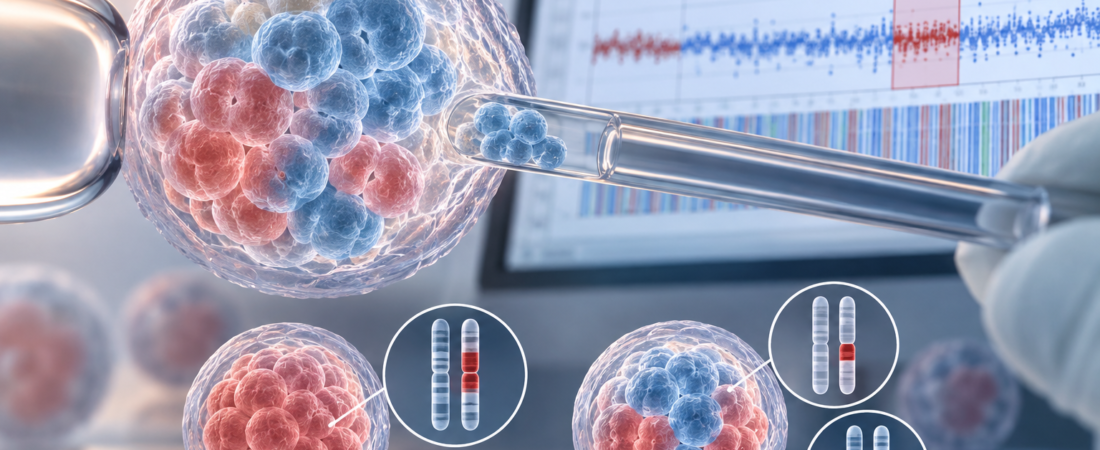
Testing three regions of 105 donated blastocysts found discordant chromosome results in 85.
These were embryos with non-mosaic segmental abnormalities—not a study showing that 81% of euploid embryos are mosaic.
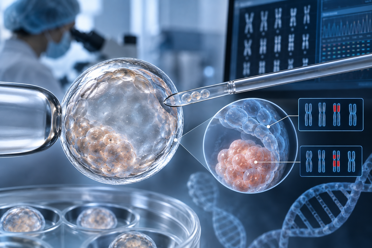
A “non-mosaic” PGT-A result may not fully describe chromosome patterns throughout an embryo, according to research on segmental aneuploidy. Among 105 blastocysts initially classified with at least one non-mosaic segmental abnormality, 85 (81%) showed different results across sampled regions.
Additional analysis changed interpretation of chromosome status in 37.1% of embryos.
The study appeared in Fertility and Sterility and was highlighted by the American Society for Reproductive Medicine in October 2026. It illustrates the limits of extrapolating from a small biopsy to an entire embryo.
Outer cells and the inner cell mass can differ
PGT-A helps inform embryo selection by testing chromosome copy-number abnormalities, usually using several outer cells from a day-5–6 blastocyst.
These trophectoderm cells mainly contribute to the placenta. The inner cell mass contains cells that develop into the fetus.
Not every cell in an embryo necessarily has the same chromosome complement. Mosaicism describes differing cell populations, potentially including cells with and without chromosome abnormalities.
An abnormality detected in the outer biopsy might not be present throughout the inner cell mass. Conversely, a biopsy without a particular abnormality cannot guarantee its absence elsewhere.
Three-region testing of 105 embryos
Balsam Al Hashimi and colleagues, including teams at the London Women's Clinic and University College London, studied 105 blastocysts donated by 86 couples.
All had at least one initial non-mosaic segmental aneuploid finding: a missing or duplicated chromosome segment, rather than an entire chromosome.
Researchers compared the original trophectoderm result with another outer-cell sample and the inner cell mass.
Results differed across the three regions in 85 embryos (81%); 20 (19%) were concordant.
ASRM's accompanying discussion reported evidence of mosaicism in 85.8% of embryos. That measure should be distinguished from the 81% figure defined by three-region discordance.
A non-mosaic classification in one biopsy therefore did not establish uniform chromosome status throughout these selected embryos.
Interpretation changed in 37.1%
Comparison across regions changed the initial interpretation in 37.1% of embryos. Some abnormalities detected initially were absent from additional samples.
Finding a normal chromosome complement in another sample does not prove that the whole embryo is normal. An abnormality could still be present elsewhere, including in the inner cell mass.
The findings do not mean PGT-A is simply wrong. They show that sampled cells may not perfectly represent the whole embryo, particularly for segmental abnormalities, which need to be distinguished from whole-chromosome findings.
Both paternal and maternal origins were identified
Paternal-origin abnormalities were identified in 52 embryos (49.5%) and maternal-origin abnormalities in 40 (38%).
Whole-chromosome numerical errors are often associated with egg formation and maternal age, but paternal origins represented a substantial component of the segmental findings here.
These numbers alone do not establish that segmental abnormalities arise mainly in sperm or are generally more often paternal than maternal.
More than half of the segmental abnormalities showed evidence of a meiotic origin, during sperm or egg formation. They therefore did not necessarily arise only during post-fertilization cell division.
In some embryos, an initial abnormality may have changed after fertilization or undergone further structural alteration. The biology can involve both gamete formation and subsequent embryo development.
What the 81% figure does not mean
These were not embryos classified as entirely euploid. They already had a segmental abnormality classified as non-mosaic.
It would be incorrect to say that 81% of embryos with normal PGT-A results are mosaic.
The study also did not transfer these embryos and measure pregnancy or birth outcomes. It cannot show that every embryo with a segmental abnormality is suitable for transfer or will result in a healthy birth.
Because PGT-A influences transfer priorities, the findings are relevant to counseling. They do not establish repeat biopsy as a universal solution; additional sampling also has limitations and potential consequences for an embryo.
The useful distinction is between what a biopsy demonstrates and what remains unknown. Decisions need to consider the type of abnormality, embryo characteristics and the patient's clinical circumstances, rather than relying on the word “non-mosaic” alone.
Study source
Balsam Al Hashimi and colleagues: “Segmental aneuploidy in human embryos is frequently mosaic with evidence of meiotic origin: insights from trophectoderm and inner cell mass analyses.” Fertility and Sterility, 2026;126(3):481–488, September issue; highlighted by ASRM in October 2026. DOI: 10.1016/j.fertnstert.2026.06.011. PMID: 42259484.
The illustration was generated using ChatGPT (OpenAI).
Related reading
Non-invasive PGT-A: what culture-medium DNA may reveal
Euploid embryo failure and the limits of a chromosome result
Copyright © 2026 THE FERTILITY NOTES. All rights reserved.