
By Seungjoo Lee
Non-invasive PGT-A in IVF could one day assess embryo chromosomes without removing cells. A new study of cell-free DNA in spent blastocyst medium suggests that fragmentomics—the patterns left when DNA breaks apart—may offer clues to embryo ploidy. This remains early research, not a replacement for embryo biopsy.
To check whether an IVF embryo has the expected number of chromosomes, laboratories currently usually remove several cells for testing. What if the liquid in which an embryo grows could provide that information without directly touching the embryo? Researchers have explored that possibility by analyzing the tiny DNA fragments left in culture medium.
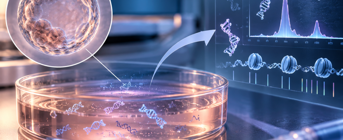
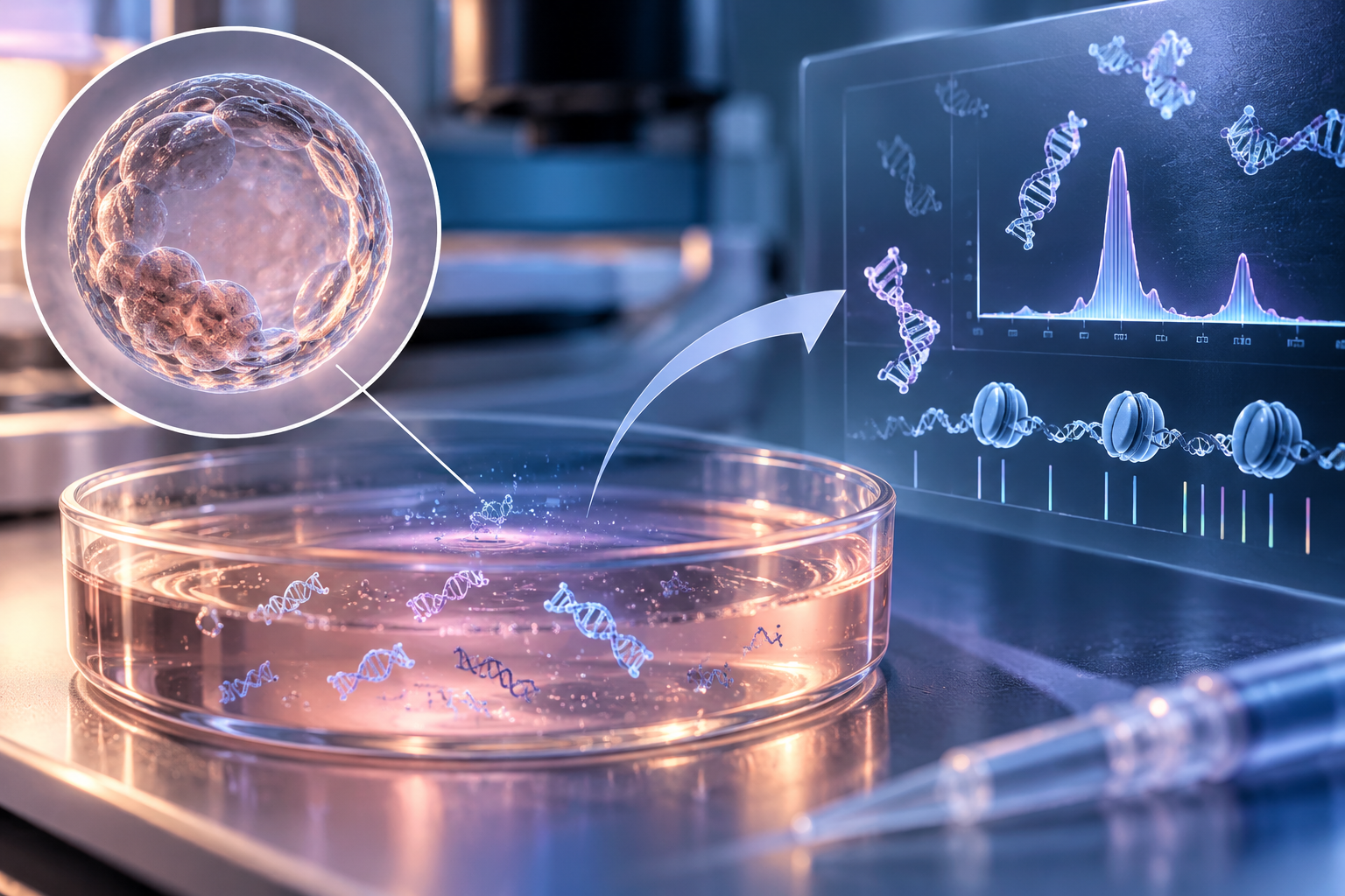
The study, published on September 22 in the Journal of Assisted Reproduction and Genetics, examined cell-free DNA (cfDNA) in spent blastocyst medium (SBM). Rather than simply measuring how much DNA was present, the team considered fragment lengths, their ends, the sequences at cleavage sites and the footprints of nucleosomes—the protein structures around which DNA is wrapped. Together, these analyses are called fragmentomics.
How PGT-A currently examines embryo chromosomes
Preimplantation genetic testing for aneuploidy (PGT-A) generally involves removing several cells from the trophectoderm, the outer blastocyst layer that contributes to the placenta. DNA from those cells is analyzed for abnormalities in chromosome number. The procedure requires skilled embryologists and specialized equipment.
The appeal of the new approach is its attempt to avoid removing embryo cells. As an embryo develops in culture, small amounts of DNA enter the surrounding medium. The investigators asked whether those molecular traces could reveal chromosomal status.
Non-invasive PGT-A using culture-medium cfDNA is not a new concept. Previous approaches amplified the small amount of DNA and compared the relative quantities associated with different chromosomes. However, culture conditions, sampling time, amplification of extremely small DNA quantities and contamination by maternal-cell DNA can affect results. Accuracy and reproducibility have not been sufficient to replace conventional PGT-A.
This study changed the question: instead of looking only at how much DNA came from each chromosome, it examined the form in which the DNA fragments remained.
150 culture-medium samples and the patterns of fragmented DNA
The researchers collected 150 spent-medium samples from 43 PGT-A cycles involving 39 patients. All oocytes were fertilized using intracytoplasmic sperm injection (ICSI), embryos were cultured individually, and medium was collected at the blastocyst stage. Six to eight trophectoderm cells were also biopsied from each corresponding embryo for conventional PGT-A, allowing comparison with the culture-medium findings.
After exclusions for library-construction failure, insufficient usable DNA data and other analysis criteria, 116 samples entered the final fragmentomic analysis: 41 euploid embryos, 28 with whole-chromosome abnormalities, 19 with complex abnormalities, nine with segmental abnormalities and 19 mosaic embryos.
The DNA had not broken randomly. Fragment lengths showed a major peak around 170 base pairs (bp), with another prominent distribution near 355 bp. Fragments shorter than 165 bp displayed a repeating pattern at approximately 10-bp intervals.
Inside cells, DNA is wrapped around nucleosomes rather than lying loose like an uncoiled thread. When cells die or break down, these structures influence where DNA is cut, potentially producing fragments with characteristic lengths and spacing.
The cfDNA floating in culture medium therefore offered more than a collection of broken molecular debris. Its lengths and cleavage patterns retained clues to biological processes in embryos and surrounding cells.
Differences near gene start sites, rather than fragment length alone
Were DNA fragments from euploid and aneuploid embryos clearly different in length? The answer was more complicated. Comparing 41 euploid embryos with 41 matched aneuploid embryos, researchers found broadly similar overall fragment-length distributions. Fragment size, end characteristics and particular end motifs did not clearly separate the two groups on their own.
A more interesting signal appeared near transcription start sites (TSSs), the locations where cells begin reading genes. The team examined the distribution of cfDNA around these sites.
Regions associated with active genes often have more open chromatin and reduced nucleosome occupancy, whereas less active regions may retain more nucleosomal structure. Looking at where cfDNA fragments remain can therefore provide indirect information about gene activity and chromatin organization in the original cells.
In this study, cfDNA coverage was greater around TSSs of genes with low expression. Coverage patterns near TSSs also differed between euploid and aneuploid embryos.
DNA footprints linked to embryo development
Genes associated with the TSS differences were involved in processes including embryo development, cytokinesis, nervous-system development and morphogenesis.
This suggests a different way of interpreting culture-medium DNA: as a record of cellular activity and chromatin structure, rather than solely the quantity of fragments from each chromosome.
The researchers also developed an exploratory model using TSS-associated cfDNA patterns. It showed potential to distinguish euploid from aneuploid embryos within the study group. The positions and fragmentation patterns of DNA left in culture medium might therefore contribute to a new kind of embryo assessment.
Why this is not yet a biopsy-free clinical PGT-A test
The results do not mean culture-medium testing can now replace PGT-A. The authors describe the model as exploratory.
First, this was a small, single-center study involving samples from 39 patients. Its predictive accuracy was not validated in a separate, large external patient population. Of the original 150 samples, 28 were excluded because of library-construction failure or inadequate unique DNA reads, illustrating the continuing difficulty of working with very small amounts of cfDNA.
Maternal DNA contamination is another unresolved issue. Not all cfDNA in the medium can necessarily be assumed to originate from the embryo: DNA from maternal cells surrounding the oocyte may be present. The investigators could not precisely quantify maternal contamination in individual samples and identified this as a limitation.
Mosaic embryos also present challenges because their chromosomal patterns and clinical implications are complex. Before clinical use, researchers would need to demonstrate reproducibility across fertility centers using different media, incubators and culture conditions.
The study is nevertheless interesting because it changes how researchers look at spent culture medium. Beyond the amount of chromosomal DNA, they can examine fragment lengths, cutting sites, nucleosomal footprints and coverage near gene start sites.
Learning about embryo chromosomes without disturbing an embryo has long been a goal of reproductive medicine. This study does not establish that the goal has been reached. It shows that a tiny drop of medium in which an embryo spent several days may contain information that has not yet been fully used.
If accuracy and reproducibility are confirmed in larger independent populations, cfDNA fragmentomics might complement morphological assessment and other non-invasive approaches. For now, it should be understood as an early investigation of whether embryo ploidy can be assessed without biopsy, rather than an established alternative to PGT-A.
Study source
Lin and colleagues, Fragmentomic features of cell-free DNA in human spent blastocyst medium and their potential for non-invasive embryo ploidy assessment, Journal of Assisted Reproduction and Genetics, September 22, 2026. Participating institutions include Nankai University School of Medicine, the University of Hong Kong–Shenzhen Hospital Reproductive Medicine Center and Shenzhen Bay Laboratory. Single-center exploratory study: 39 patients, 43 cycles, 150 collected samples and 116 final analyzed samples. DOI: 10.1007/s10815-026-04033-7; PMID 42771117.
Related reading
- The embryologist’s role and IVF laboratory safety
- PPOS and euploid embryos: randomized trial evidence
Images were created using generative AI (ChatGPT, OpenAI) for illustration and do not depict actual patients.
Copyright © 2026 Fertility Information TokTok | THE FERTILITY NOTES. All rights reserved. Unauthorized republication, reproduction, redistribution, scraping, AI training and data use of articles, photographs, images and all other content are strictly prohibited. Violations may result in legal liability.