
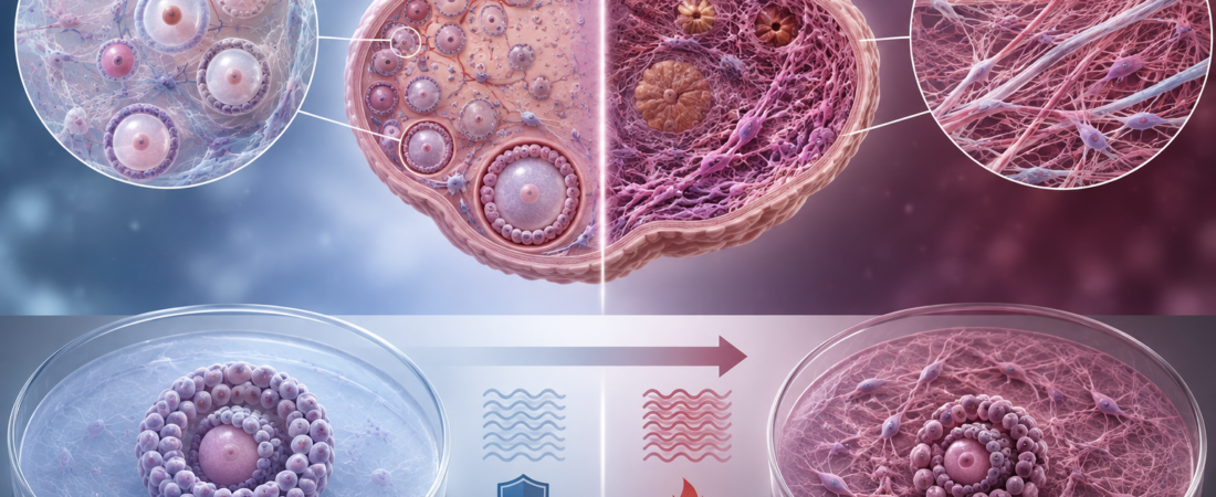
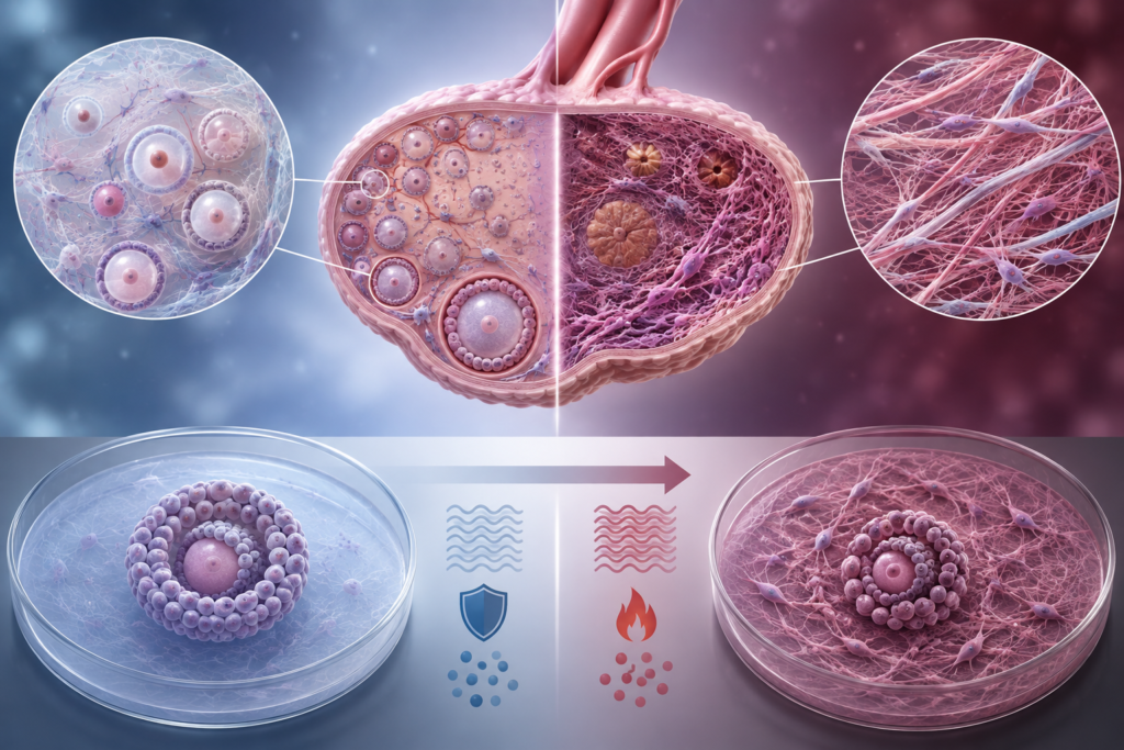
- Collagen Accumulates in Aging Human Ovarian Cortex: Structural fibrosis and directional realignment of fibrillar collagen confirmed in post-menopausal tissue
- Stiff Mechanical Environments Trigger Fibro-Inflammatory Cascades: Culturing healthy follicles in rigid hydrogels impairs growth, reduces estradiol secretion, and degrades oocyte quality
- Disrupted Oocyte-Somatic Crosstalk: Tissue stiffness decreases transzonal projections (TZPs), severing physical communication between granulosa cells and the oocyte
- Shifting Research Focus: Ovarian aging expands beyond numerical “egg depletion” toward biomechanical degradation of the ovarian niche
Age-related reproductive decline in women has long been defined by two primary metrics: the quantitative depletion of the primordial follicle pool and the qualitative decline in oocyte nuclear and mitochondrial competence. However, emerging research introduces a critical third dimension: the physical and mechanical aging of the ovarian tissue itself.
Rather than serving as an inert biological container, the extracellular matrix (ECM) of the aging ovary undergoes progressive fibrosis and biomechanical stiffening, creating a hostile physical microenvironment that directly impairs follicular development and oocyte health.
Collagen Deposition in the Human Ovarian Cortex
A study published in Molecular Reproduction and Development (April 2026) by researchers at the University of Ottawa demonstrated structural remodeling in human ovarian tissue across reproductive stages.
Analyzing pre- and post-menopausal ovarian samples, the researchers mapped collagen architecture across the outer cortex (where primordial and early-stage follicles reside) and the inner medulla:
- Selective Cortical Fibrosis: Fibrillar collagen was significantly elevated and concentrated specifically within the ovarian cortex of post-menopausal tissue, whereas the medulla showed no equivalent age-related change.
- Structural Realignment: Beyond an increase in collagen volume, aging ovaries exhibited thicker, densely packed collagen bundles realigned with uniform directionality.
Because primordial and primary follicles reside in the cortex throughout their decades-long dormancy, this progressive remodeling means developing follicles must navigate a drastically stiffer and denser extracellular matrix.
Mechanical Rigidity as an Independent Driver of Follicular Decline
To investigate whether ovarian stiffness is merely a benign biomarker of aging or an active driver of follicular dysfunction, a study published in Reproduction (January 2026) by Washington University School of Medicine isolated mechanical rigidity as a single experimental variable.
Researchers cultured secondary follicles from young, healthy mice inside 3D alginate hydrogels calibrated to mimic either a soft (young) or stiff (aged) ovarian microenvironment:
- Acute Fibro-Inflammatory Activation: Within hours of exposure to the rigid matrix, follicles upregulated the expression of genes associated with sterile inflammation and pathological ECM remodeling.
- Impaired Growth & Endocrine Secretion: Long-term culture in stiff matrices resulted in reduced follicular survival rates, lower granulosa cell viability, and suppressed estradiol production.
- Compromised Oocyte Competence: Oocytes retrieved from follicles grown in stiff matrices exhibited significantly reduced maturation rates and poorer morphological quality.
Crucially, because this experiment utilized healthy follicles from young donors, the observed dysfunction was triggered entirely by the physical stiffness of the surrounding matrix, demonstrating that biomechanical forces alone can induce an aged phenotype.
Severing the Transzonal Projections (TZPs)
The study identified a critical structural mechanism explaining this loss of oocyte quality: the loss of transzonal projections (TZPs).
An oocyte depends entirely on metabolic and biochemical support delivered by surrounding granulosa cells. This communication occurs through TZPs—specialized, microscopic filopodia-like cellular bridges that extend from granulosa cells, penetrate the zona pellucida, and form gap junctions with the oolemma.
In the stiff matrix environment:
- The density of intact TZPs dropped significantly.
- Mechanical compression from the rigid ECM disrupted the delicate physical contacts between the somatic granulosa cells and the growing oocyte, impairing nutrient transport, cyclic nucleotide delivery, and metabolic cooperation.
Modulating Matrix Stiffness: The Role of IL-11
Parallel findings published in Nature Aging (July 2026) utilized atomic force microscopy (AFM) to directly measure the nanoscale elasticity of human and mammalian ovaries, confirming a progressive, age-dependent increase in cortical Young’s modulus (rigidity).
The researchers identified the pro-inflammatory cytokine Interleukin-11 (IL-11) as a central mediator stimulating stromal fibroblasts to drive collagen deposition and matrix stiffening:
- Targeted Inhibition: In preclinical rodent models, blocking IL-11 signaling attenuated fibrotic remodeling, normalized ovarian matrix stiffness, and extended reproductive longevity.
- While IL-11-targeted therapies remain in preclinical stages and are not yet established human treatments, they provide proof-of-concept that ovarian fibrosis is an active biological pathway amenable to pharmacological modulation.
Reframing Ovarian Reserve: Beyond Endocrine Numbers
Standard clinical assessments—such as Anti-Müllerian Hormone (AMH), basal Follicle-Stimulating Hormone (FSH), and Antral Follicle Count (AFC)—gauge the remaining quantity of follicles. However, they provide no information regarding the degree of stromal fibrosis or physical stiffness within the ovarian cortex.
| Clinical Parameter | Traditional Focus | Emerging Biomechanical Paradigm |
| Primary Metric | Oocyte quantity (AMH, AFC) & ploidy | ECM composition, collagen density, and tissue elasticity |
| Mechanistic View | Passive depletion / intrinsic oocyte aging | Active microenvironmental degradation & fibrosis |
| Cellular Target | Oocyte spindle & mitochondria | Granulosa-oocyte communication (TZPs) & stromal fibroblasts |
| Therapeutic Horizon | IVF stimulation & embryo selection | ECM remodeling, anti-fibrotic & anti-inflammatory agents |
Clinical Context and Limitations
While these biomechanical insights are transformative for reproductive biology, key clinical caveats remain:
- Translational Gap: The functional assays demonstrating that stiffness impairs oocyte quality were primarily performed in rodent 3D in vitro follicle culture systems; human clinical trials reversing ovarian stiffness are not yet available.
- Multifactorial Aging: Ovarian senescence involves complex, concurrent mechanisms—including meiotic cohesin deterioration, mitochondrial DNA deletions, oxidative damage, and systemic endocrine changes. Fibrosis represents an important contributing axis, but not the sole cause of reproductive aging.
The scientific paradigm of ovarian aging is shifting: moving from viewing the ovary simply as a dwindling warehouse of eggs to understanding it as an interconnected biomechanical niche where the “house” ages alongside its inhabitants.
Medical Sources & Literature References
- Journal: Molecular Reproduction and Development (April 2026) — Ovarian Fibrosis Through Comparative Analysis of Collagen Architecture (University of Ottawa)
- Journal: Reproduction (January 2026) — Increased stiffness mimicking ovarian aging induces a fibroinflammatory response in follicles and impairs oocyte quality (Washington University School of Medicine)
- Journal: Nature Aging (July 2026) — Modulating IL-11-dependent matrix stiffness to delay ovarian aging
※ This article was synthesized based on peer-reviewed biomechanical and reproductive biology studies published in Molecular Reproduction and Development, Reproduction, and Nature Aging (2026). It does not replace individualized clinical diagnosis or medical care, and specific treatment decisions should always be made in consultation with a qualified reproductive specialist.
※ The images associated with this article were generated using generative AI (ChatGPT, OpenAI) as illustrative visual references and do not depict real individuals.