
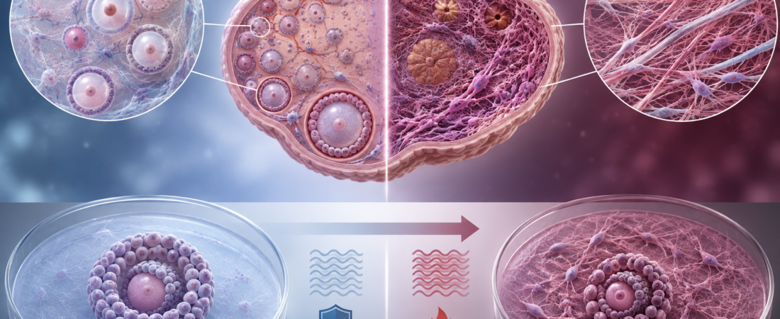
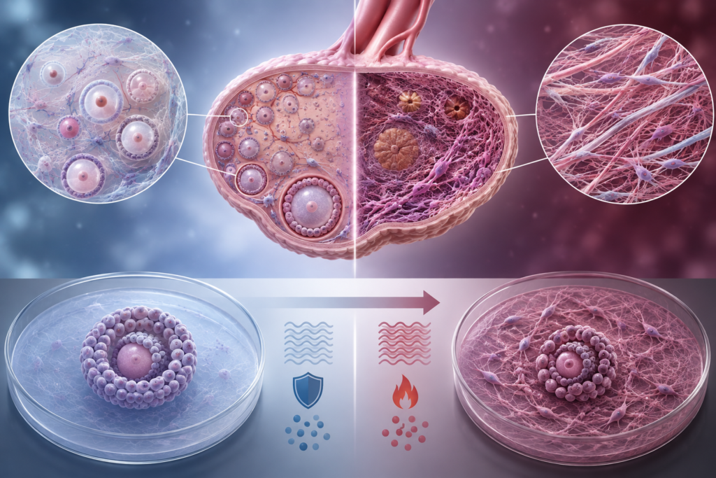
- Capturing Granulosa Cell Metabolic Shifts After Age 35: Reduced mitochondrial oxidative phosphorylation and increased compensatory reliance on glycolysis
- 25 Metabolites Significantly Altered in GCs of IVF Patients: New insights into the molecular bioenergetics of ovarian aging
- Expanding from Oocyte-Centric Aging to the Follicular Microenvironment: A growing body of evidence shows the cellular nursery ages alongside the egg
As maternal age advances, the quantity of remaining oocytes declines and the rate of chromosomal aneuploidy rises. Historically, ovarian aging has been framed almost exclusively around these two hallmarks. However, recent research looks deeper into the follicle, demonstrating that the cellular energy infrastructure of the somatic cells surrounding and nourishing the egg undergoes profound age-related deterioration.
Within the ovarian follicle, granulosa cells (GCs) are far more than a passive protective shield. They actively synthesize steroid hormones, drive follicular growth, and supply essential metabolic substrates and nutrients to the developing oocyte, shaping the biochemical microenvironment required for cytoplasmic and nuclear maturation.
A study published in Frontiers in Endocrinology analyzed how the energy metabolism of these granulosa cells shifts with maternal age. Researchers conducted targeted metabolomic profiling on human mural granulosa cells collected from women undergoing In Vitro Fertilization (IVF), comparing younger women (ages 21–34) against women of advanced reproductive age (ages 35–42), with 10 biological replicates per cohort in the core metabolomics validation.
Altered Energy Generation Within the Follicle
The metabolomic profiling revealed distinct age-associated divergences:
- 25 Statistically Significant Metabolites: While 32 metabolites initially differed between age groups, 25 metabolites retained statistical significance after rigorous false discovery rate (FDR) correction (17 upregulated and 8 downregulated in the advanced age cohort).
- Key Pathway Clustering: Metabolic alterations were heavily concentrated across three core bioenergetic axes: Oxidative Phosphorylation (OXPHOS), Glycolysis, and the mitochondrial Tricarboxylic Acid (TCA) cycle.
Compared to younger granulosa cells, GCs from women aged 35–42 exhibited significant elevations in metabolites involved in glycolysis—the rapid, anaerobic breakdown of glucose. Conversely, metabolite flux and efficiency through mitochondrial oxidative phosphorylation and the TCA cycle were notably attenuated.
This represents a fundamental shift in cellular energy strategy: metabolic reprogramming.
Under physiological conditions, healthy mitochondria utilize pyruvate and oxygen via the TCA cycle and electron transport chain to generate Adenosine Triphosphate (ATP) with high efficiency. When mitochondrial integrity and respiratory chain function decline, the cell adapts by upregulating glycolysis to meet baseline bioenergetic demands, compensating for compromised mitochondrial output.
Metabolic Shift Rather Than Pure Energy Cessation
The findings do not imply that granulosa cells in older follicles stop producing energy altogether; rather, the pathway distribution of energy production is altered.
To functionally validate these human metabolomic findings, the researchers conducted in vitro assays using the human granulosa-like cell line (KGN) subjected to oxidative stress to model cellular aging:
- Oxygen Consumption Rate (OCR): Respiration and mitochondrial oxygen consumption dropped markedly in senescent GCs.
- Extracellular Acidification Rate (ECAR) & Lactate: Acidification and lactate secretion increased, confirming elevated glycolytic flux.
- Mitochondrial Reactive Oxygen Species (mtROS): Intracellular and mitochondrial oxidative stress markers rose significantly.
While mitochondria act as the primary, high-yield power plant of the cell, glycolysis serves as a lower-yield, emergency backup generator. Heavy reliance on glycolysis indicates that granulosa cells are operating under severe mitochondrial bioenergetic strain.
From “Egg Aging” to “Follicular Aging”
This study is significant because it broadens the paradigm of reproductive aging from an isolated oocyte defect to an ecosystem-wide failure of the follicular microenvironment.
An oocyte does not develop in isolation. It is metabolically coupled with its surrounding granulosa and cumulus cells via transzonal projections and gap junctions. Because the mammalian oocyte has limited intrinsic capacity to utilize glucose directly, it relies heavily on surrounding somatic cells to process glucose and import downstream metabolites (such as pyruvate) for its own maturation.
Consequently, when granulosa cell mitochondria falter and their metabolic output shifts, the oocyte is deprived of optimal metabolic support. This suggests that poor oocyte quality in older women is not driven solely by nuclear chromosomal errors, but also by the progressive aging and bioenergetic exhaustion of the follicular niche that nurtures it.
Limitations and Clinical Interpretation
Several considerations must be kept in mind when interpreting the data:
- Sample Size: The targeted metabolomics analysis was conducted on a relatively small cohort of 20 patients (10 young vs. 10 older).
- Clinical Setting: All samples originated from patients undergoing IVF for clinical subfertility, meaning findings should be confirmed in broader fertile populations.
- Continuous Biological Process: The 35-year-old threshold represents a standard clinical stratification benchmark; reproductive aging is a continuous, individualized biological spectrum rather than an abrupt metabolic cliff.
- Preclinical Stage: Identifying metabolic reprogramming identifies potential molecular targets, but this study was not an interventional trial demonstrating that altering these metabolic pathways will directly increase clinical live birth rates.
Reframing the Reproductive Aging Narrative
Historically, clinical discussions around age-related fertility decline have centered on two metrics: How many eggs remain (AMH/AFC)? and Are their chromosomes normal (euploidy)?
Recent discoveries introduce a critical third biological question:
“In what bioenergetic and metabolic environment are those remaining eggs developing?”
Ovarian aging is more than a passive depletion of oocyte inventory. It is an active, multi-tissue aging process where the supporting cellular machinery and mitochondrial power plants within the follicle undergo functional decline. Research is steadily moving toward understanding—and potentially supporting—the somatic microenvironment that sustains oocyte competence.
Medical Source & Study Information
- Journal: Frontiers in Endocrinology (Published February 2026)
- Study Title: Age-related mitochondrial energy metabolism reprogramming occurs in granulosa cells during ovarian aging
- DOI: 10.3389/fendo.2026.1726339
※ This article was synthesized based on research published in Frontiers in Endocrinology (February 2026) and literature in reproductive endocrinology. It does not replace individualized clinical diagnosis or medical care, and specific medical decisions should always be made in consultation with a qualified reproductive specialist.
※ The images associated with this article were generated using generative AI (ChatGPT, OpenAI) as illustrative visual references and do not depict real individuals.
Want to explore targeted adjuvant therapies studied to support granulosa cell mitochondrial function (like CoQ10 or resveratrol)?예