
By Seungjoo Lee
Intrauterine fluid during frozen embryo transfer (FET) preparation was associated with a lower live birth rate in a study of 160 women. The amount of fluid appeared relevant, but the exploratory 2.55-mm threshold is not a universal rule for proceeding with or canceling embryo transfer.
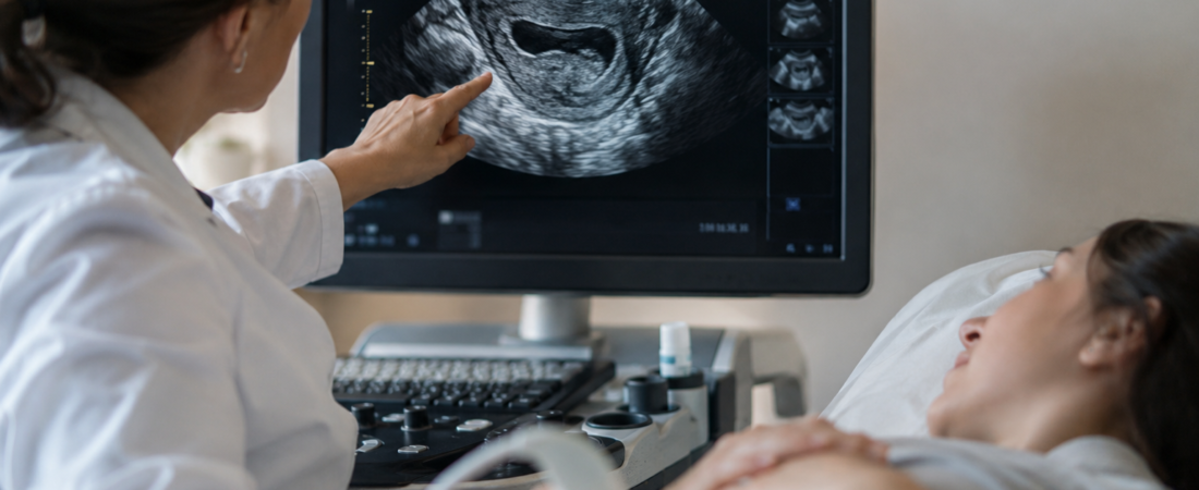
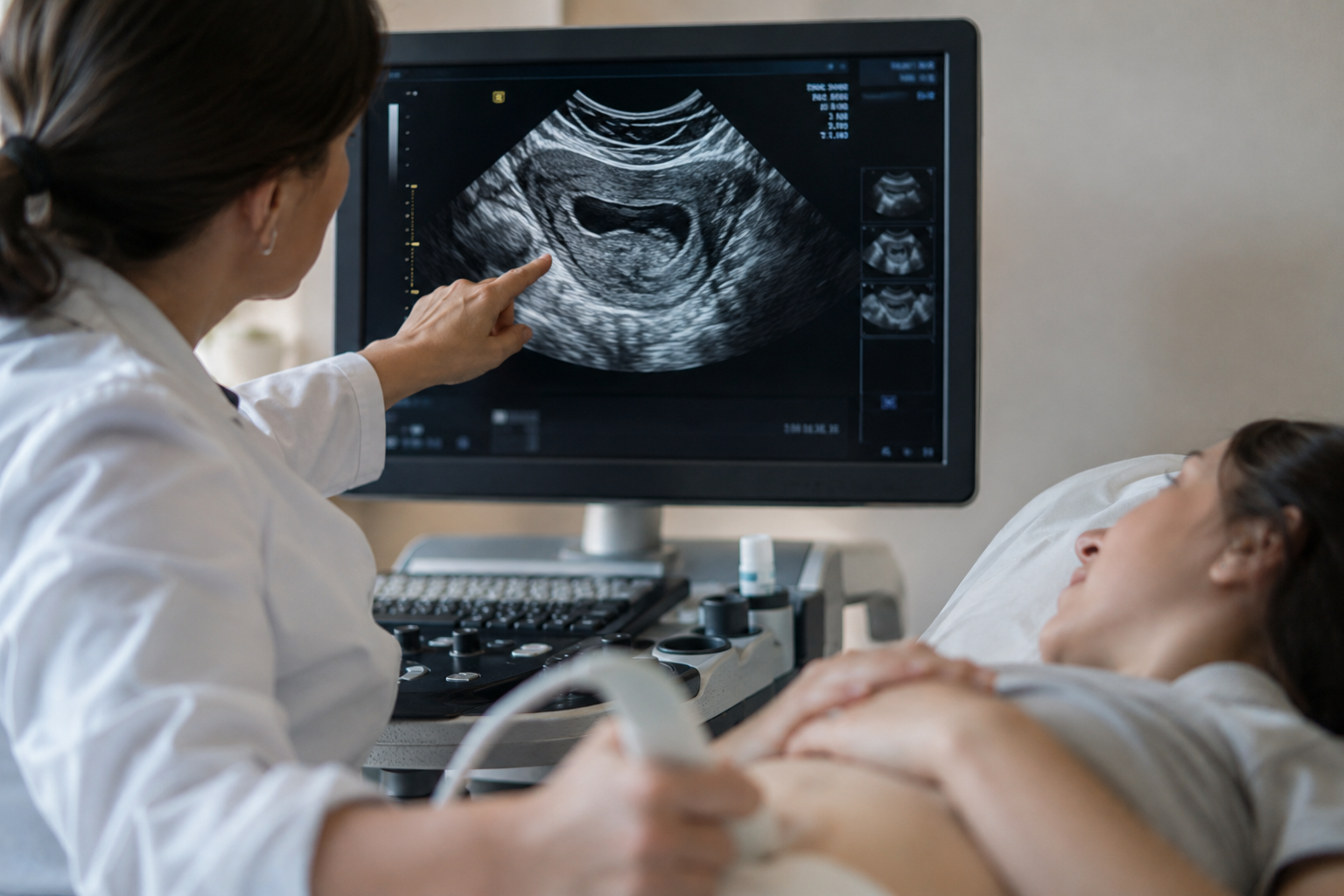
“There is a little fluid inside the uterus.” Hearing this during an ultrasound can be unsettling for someone preparing the endometrium to receive a hard-won frozen embryo. Can an embryo implant if fluid is present, and can transfer go ahead?
A recent study found that larger collections of intrauterine fluid (IUF) were associated with lower odds of live birth. Researchers emphasized that assessment should consider the amount, cause and persistence of fluid, as well as the endometrium, rather than simply whether fluid is present.
What does fluid in the uterine cavity mean?
Fluid seen on ultrasound is generally within the cavity between the endometrial surfaces, where an embryo is transferred and implants—not within the uterine muscle itself.
Causes vary. This study grouped fluid by isthmocele, intrauterine adhesions (synechiae) and idiopathic cases in which no clear cause was identified.
An isthmocele is a pouch-like defect in a previous cesarean-section scar. Blood or mucus can collect there and enter the cavity. Intrauterine adhesions occur when the inner uterine surfaces adhere to one another following events such as curettage, intrauterine surgery or inflammation.
Hydrosalpinx is another clinically important consideration. Fluid trapped in a blocked fallopian tube can flow back toward the uterus and create an unfavorable implantation environment. Similar-looking fluid on ultrasound can therefore have different causes and implications.
Live birth rates: 27.7% with fluid versus 42.9% without
The investigators compared 83 women with fluid detected during FET preparation with 77 women preparing for FET during the same period without fluid, analyzing 160 women in total.
Fluid size was measured by transvaginal ultrasound at the maximum anteroposterior diameter. Outcomes were examined in relation to fluid cause, treatment and dimensions.
The live birth rate was 27.7% in women with IUF and 42.9% in controls, a statistically significant difference of approximately 15 percentage points. Yet the central question extended beyond the presence of fluid: the amount of fluid also appeared to matter.
Greater fluid dimensions were associated with lower live birth odds
After adjustment for other factors, each 1-mm increase in fluid dimension was associated with lower live birth odds, with an adjusted odds ratio of 0.29. This is an odds-based association, not an individual probability estimate.
Exploratory analyses identified 3.10 mm as a value discriminating positive pregnancy tests and 2.55 mm as a value discriminating live birth outcomes in this study population.
These are not clinical rules saying that transfer should proceed below a threshold and be canceled above it. They are statistical cutoffs identified within a relatively small study and require confirmation in larger populations. A measurement of 2.5 or 3 mm alone cannot determine whether a particular patient should undergo transfer.
Treated and untreated patients had different outcomes
Among women with IUF, the live birth rate was 38.6% in those receiving medical or surgical treatment and 15.4% in those who did not. The odds of live birth were 3.46 times higher in the treated group.
This finding also needs cautious interpretation. Women were not randomly assigned to treatment or no treatment. Causes of fluid, selection for treatment and the treatments received could differ substantially.
It does not mean that treating any fluid collection triples an individual’s chance of live birth. It suggests that identifying the cause and treating appropriately selected patients may be important.
Adhesions were associated with a less favorable endometrium
Pregnancy outcomes did not differ significantly across the isthmocele, synechiae and idiopathic subgroups. However, the adhesion subgroup had the thinnest endometrium and a lower frequency of a trilaminar pattern: 38.5%, compared with 69.7% in the comparison group.
A trilaminar endometrium appears as a three-line pattern on ultrasound and is assessed alongside thickness during transfer preparation.
This helps explain why simply removing visible fluid may not resolve the entire problem. Underlying adhesions or a cesarean-scar defect may also affect the endometrial environment.
Cause, amount and persistence matter together
A single observation of fluid during FET preparation does not establish that pregnancy is unlikely. A small collection may appear temporarily and then resolve.
Repeated, increasing or persistent fluid near transfer warrants a different assessment. Clinicians may need to investigate adhesions, scar defects, hydrosalpinx or other conditions that require treatment.
Combining fluid size, cause and persistence with endometrial thickness and appearance provides more information than a single measurement or a yes/no finding. A small transient collection is not necessarily equivalent to a larger persistent collection.
When fluid is seen before FET, useful questions include where it came from, how much is present and whether it remains on the next scan. The study suggests an independent association between fluid dimensions and live birth, but does not establish a cancellation threshold applicable to every patient. The whole clinical picture matters.
Study source and limitations
Intrauterine fluid during frozen embryo transfer preparation: Impact of etiology, treatment, and fluid dimension on pregnancy outcomes, European Journal of Obstetrics & Gynecology and Reproductive Biology, online September 23, 2026. Investigators included researchers at Acıbadem Izmir Hospital, Acıbadem Maslak Hospital and Acıbadem University, Türkiye. Observational comparison of 83 women with IUF and 77 controls; treatment was not randomized and thresholds were exploratory. DOI: 10.1016/j.ejogrb.2026.115454; PMID 42800372.
This article provides research information and does not replace clinical decisions about proceeding with or canceling FET, or whether treatment is appropriate.
Related reading
Images were created using generative AI (ChatGPT, OpenAI) for illustration and do not depict actual patients.
Copyright © 2026 Fertility Information TokTok | THE FERTILITY NOTES. All rights reserved. Unauthorized republication, reproduction, redistribution, scraping, AI training and data use of articles, photographs, images and all other content are strictly prohibited. Violations may result in legal liability.