
Reading ‘Childbirth Potential’ Through Embryo Metabolites 173 Metabolites Detected in Spent Culture Medium… 10 Differed Based on Live Birth Outcomes Live Birth Discrimination Model Achieves AUC of 0.834… The Promise of ‘Non-Invasive Selection’ Without Touching Embryos
Moving beyond the traditional practice of selecting high-quality embryos based solely on visual morphology under a microscope, recent research explores assessing childbirth potential through the “metabolic footprints” embryos leave behind in their culture medium.
In a study published in the international journal Reproductive BioMedicine Online (RBMO), researchers analyzed 70 spent embryo culture medium samples from patients who underwent single blastocyst transfers using mass spectrometry.
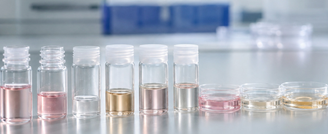
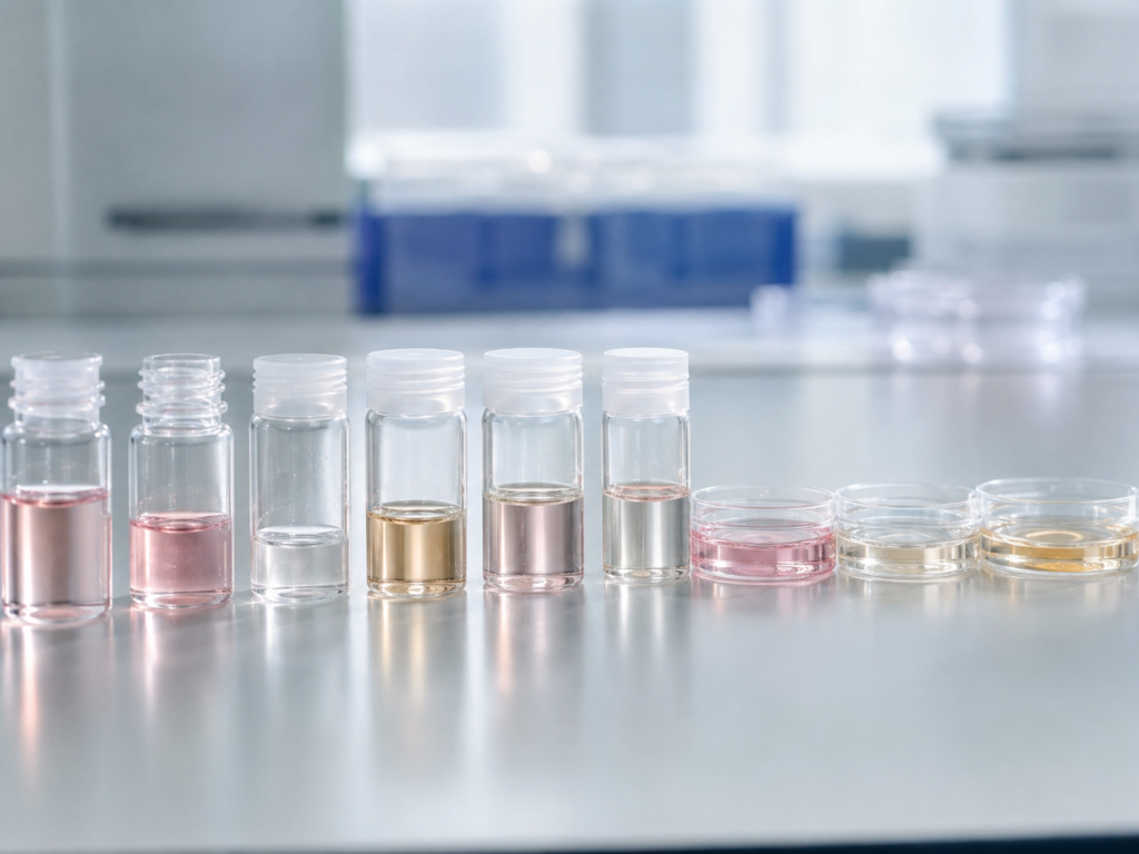
This medium—known as spent embryo culture medium (SECM)—is the residual fluid left behind after an embryo consumes nutrients and excretes metabolic byproducts throughout its growth.
Detecting Metabolic Signatures of Live Birth
The analysis detected a total of 173 metabolites within the culture media:
- 6 metabolites exhibited significant differences based on whether clinical pregnancy was achieved.
- 10 metabolites demonstrated distinct differences correlating with actual live birth outcomes.
Notably, when researchers constructed a predictive model using the metabolic profiles of the culture media to discriminate live birth outcomes, it achieved an Area Under the Curve (AUC) of 0.834. AUC serves as a metric indicating how accurately a predictive model differentiates between two groups (where 0.5 represents random chance and values approaching 1 reflect superior discriminatory power).
This does not imply that testing culture media can already flawlessly predict which embryo will lead to a live birth. However, this study commands attention because it demonstrates the viability of extracting chemical footprints left behind during development without biopsying or touching the embryo itself.
Moving Beyond Morphology and Invasive Biopsy
Standard IVF currently relies heavily on morphological grading, assessing criteria such as blastocyst expansion, the inner cell mass (ICM), and the trophectoderm (TE). When deemed necessary, clinics perform preimplantation genetic testing (PGT), which requires biopsying a small cluster of cells from the embryo to screen chromosomes.
Spent medium metabolomic profiling takes a fundamentally different approach. Rather than evaluating outward appearance, it asks: “What did the embryo consume, and what did it release into the droplet?”
Even among embryos that appear morphologically identical under a microscope, variations in metabolic activity can exist. These metabolic discrepancies may offer novel predictive insight into implantation capacity, clinical pregnancy, and ultimate live birth potential.
Limitations and the Road Ahead
This study carries clear limitations as a pilot study analyzing 70 culture medium samples. The findings must be replicated in trials with substantially larger patient cohorts and embryo volumes. Furthermore, future validation must account for variations across different culture media brands, incubation environments, and culturing durations.
Even so, it is noteworthy that IVF embryo selection research is actively expanding its scope from the “visible embryo” to the “traces an embryo leaves behind.”
Future embryo assessment may evolve beyond simply finding the most aesthetically symmetrical embryo under a microscope to jointly decoding the subtle metabolic signals an embryo secretes into its culture droplet over several days of development.
※ This article was synthesized based on research published in the international journal Reproductive BioMedicine Online (RBMO). It does not replace individual medical diagnosis or clinical care, and specific medical decisions must be made through consultation with a qualified specialist.
※ The images used in this article were generated using generative AI (ChatGPT, OpenAI) as visual references to aid understanding and do not depict real individuals.