
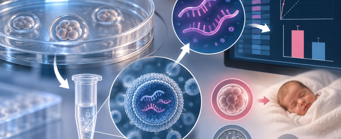
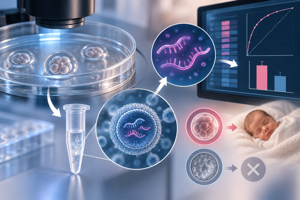
- Profiling exosomal microRNA (miRNA) in spent culture medium (SCM):
hsa-miR-375-3psignificantly elevated in culture droplets of embryos resulting in live birth - Diagnostic performance: $\text{AUC} = 0.763$, sensitivity 85%, specificity 70%—demonstrating the potential for non-invasive embryo assessment without mechanical biopsy
- Functional mouse assays confirm active biological role: suppressing
miR-375-3psignificantly impaired embryonic progression at the 4-to-8 cell cleavage stage - Single-center pilot study ($n = 67$): Serves as a potential complementary biomarker for developmental competence, not an immediate replacement for PGT-A
Embryo selection in In Vitro Fertilization (IVF) is expanding beyond “inspecting the embryo itself” to “decoding the molecular footprints it leaves behind.”
A study published in Reproductive BioMedicine Online (RBMO) explored whether analyzing trace molecules within the droplets of fluid left after culturing human embryos can predict which embryo is most likely to result in a live birth.
The central candidate identified was a microRNA designated hsa-miR-375-3p. While this non-invasive methodology remains in early research stages and is not yet ready for routine clinical adoption, it represents a step toward evaluating an embryo’s developmental competence without the cellular stress of an invasive biopsy.
Inspecting the Secretome Rather Than Breaching the Embryo
Standard laboratory assessment of embryo quality relies primarily on high-resolution optical microscopy to grade cell symmetry, fragmentation, and blastocyst expansion. More recently, time-lapse imaging (TLI) paired with artificial intelligence (AI) algorithms has enabled continuous tracking of dynamic cell division intervals (morphokinetics).
Conversely, Preimplantation Genetic Testing for Aneuploidy (PGT-A) takes an invasive approach: using a laser to breach the protective zona pellucida and aspirating 5 to 10 cells from the trophectoderm (the precursor to the placenta) for chromosomal copy-number analysis.
This study evaluated an entirely non-invasive paradigm: spent culture medium (SCM) profiling.
Throughout in vitro culture, an embryo metabolizes nutrients and actively secretes signaling molecules, metabolites, and nucleic acids into the surrounding micro-droplet of culture medium. Rather than touching or biopsying the embryonic cells, researchers harvest the fluid left behind after the embryo is removed for transfer or cryopreservation, analyzing its molecular “secretome.”
Exosomes: Microscopic Cargo Shuttles in the Culture Droplet
The investigators focused their analysis specifically on embryonic exosomes isolated from the spent culture medium.
Exosomes are nanoscale extracellular vesicles (EVs) secreted by living cells that package functional biological cargo, including proteins, lipids, and non-coding regulatory RNAs. They function essentially as molecular messaging capsules operating between cells:
- Small Non-Coding RNA Profiling: The researchers extracted exosomes from the droplet fluid and profiled their non-coding RNA composition, identifying 51 microRNAs (miRNAs), 547 PIWI-interacting RNAs (piRNAs), and 39 transfer RNA fragments (tRFs).
- Biomarker Identification: Several molecules exhibited divergent expression profiles between embryos that achieved live birth and those that failed. Most notably,
hsa-miR-375-3p(andmiR-215-5p) were significantly enriched in the exosomes of embryos that progressed to successful delivery. - Predictive Performance: Receiver Operating Characteristic (ROC) curve analysis of
miR-375-3pyielded an Area Under the Curve ($\text{AUC}$) of 0.763, with a sensitivity of 85% and a specificity of 70%.
An $\text{AUC}$ of 0.763 indicates moderate-to-good discriminatory capability in distinguishing live-birth from non-live-birth outcomes. However, it also means miR-375-3p is an exploratory candidate biomarker rather than an absolute standalone diagnostic test.
Comparison of Clinical Embryo Evaluation Modalities
| Evaluation Modality | Target Analyzed | Invasiveness | Primary Clinical Objective | Inherent Limitations |
| Standard Morphology | Visual shape, fragmentation, blastocyst expansion | Non-invasive | Baseline embryo grading | Subjective; poor correlation with chromosomal ploidy |
| Time-Lapse Imaging (AI) | Dynamic cleavage timing & division kinetics | Non-invasive | Ranking embryos by kinetic competence | High equipment cost; cannot identify chromosomal aneuploidy |
| Trophectoderm PGT-A | 5–10 cells biopsied from trophectoderm | Invasive | Screening numerical chromosomal aneuploidy | Requires blastocyst biopsy; risk of cell damage; mosaicism ambiguity |
| Spent Medium Exosomal miRNA (Emerging) | Secreted exosomal cargo (miR-375-3p) in SCM | Non-invasive | Assessing intrinsic developmental competence | Experimental; requires standardized isolation protocols & larger validation |
Functional Validation: Active Role in Early Embryonic Cleavage
The research team went beyond establishing a statistical correlation. To determine whether miR-375-3p plays an active biological role during early development, they conducted functional loss-of-function experiments in murine embryos.
By microinjecting targeted miR-375-3p inhibitors into early cleavage-stage mouse embryos, the researchers observed a statistically significant reduction in the rate of 8-cell embryo formation ($P = 0.0358$).
This functional assay indicates that miR-375-3p is actively involved in regulating the critical transition from the 4-cell to the 8-cell stage—a window that coincides with the activation of the embryonic genome (the maternal-to-zygotic transition). Its presence in the culture medium appears to reflect genuine developmental viability rather than non-specific cellular leakage or random debris.
Can Spent Culture Medium Replace PGT-A?
Extrapolating these findings to suggest that clinicians can immediately discard embryo biopsies in favor of a single drop of culture fluid would be premature:
- Cohort Size and Design: The study analyzed a single-center cohort of 67 Day-3 embryos (all morphologically top-grade Grade 1 cleavage embryos from single-embryo transfers: 34 resulting in live birth, 33 in non-birth). Whether these findings hold across lower-grade embryos, Day-5/6 blastocysts, older maternal demographics, or diverse clinical etiologies remains unproven.
- Distinct Biological Questions: PGT-A specifically evaluates whole-chromosome numerical copy numbers (aneuploidy). Exosomal miRNA analysis assesses metabolic and transcriptomic vitality. An embryo can be chromosomally euploid yet lack the metabolic vigor to sustain development, or conversely, an aneuploid embryo might transiently secrete normal levels of regulatory miRNAs. Therefore, SCM testing cannot simply substitute for genetic ploidy testing.
- Background Contamination: Commercial embryo culture media contain complex protein supplements (such as human serum albumin) that can introduce exogenous vesicles, requiring rigorous isolation protocols to avoid false-positive reads.
Rather than replacing PGT-A, the true near-term value of exosomal miRNA profiling lies in complementing non-invasive selection. In scenarios where multiple embryos appear identical under the microscope and patients choose not to undergo invasive genetic biopsy, secretomic biomarkers could provide an objective metric to rank which embryo should be prioritized for intrauterine transfer.
From Visual Inspection to Molecular Decoding
Embryo assessment in reproductive medicine continues to evolve. What began decades ago as a simple static look through a microscope has advanced through continuous time-lapse monitoring and molecular genetic testing.
This study outlines the next phase of that progression: decoding the active communication signals an embryo releases into its immediate surroundings.
While larger prospective multicenter trials are needed before clinical assays can be deployed in IVF laboratories, the biological premise is clear. An embryo’s culture droplet is not merely disposable fluid—it contains a molecular record of its developmental health. The ongoing challenge is learning how to read those signals with clinical precision.
Medical Source & Study Reference
- Journal: Reproductive BioMedicine Online (RBMO), Paper ID: 105477 (Published online January 16, 2026).
- Study Title: hsa-miR-375-3p in embryo culture medium exosomes as a preimplantation non-invasive biomarker for predicting live birth after IVF
- Lead Institutions: Xinjiang University; Xinjiang Jiayin Hospital, Urumqi, China.
- Study Cohort: 67 single Day-3 Grade 1 embryo transfers (34 live births vs. 33 non-live births) with exosomal small non-coding RNA profiling and murine functional knockdown validation.
- Identifiers: DOI: 10.1016/j.rbmo.2026.105477 | PMID: 42400996
※ This article provides general scientific and medical information and does not replace individualized clinical diagnosis or embryo transfer decisions. Embryo prioritization must be determined collaboratively by the attending reproductive endocrinologist and embryology team, incorporating morphological grading, patient clinical history, and validated diagnostic testing. Exosomal miRNA analysis of spent culture medium remains an experimental technology and is not an established clinical standard of care.
※ The images associated with this article were generated using generative AI (ChatGPT, OpenAI) as illustrative visual references and do not depict real individuals.
※ Copyright © 2026 THE FERTILITY NOTES. All rights reserved. Unauthorized reproduction, duplication, distribution, scraping, AI training, or data utilization of all content, including articles, photos, and images, is strictly prohibited and may result in legal liability.