

- Multicenter study across 8 fertility centers: Live birth chances significantly lower in women with adenomyosis
- Disparities remain even with donor eggs and euploid embryos: The uterus itself emerges as an independent variable
- Poorer pregnancy outcomes linked to intrinsic adenomyosis near the endometrium and severe lesions
In In Vitro Fertilization (IVF), there are times when patients secure high-grade embryos, only to face implantation failure or pregnancy loss after a positive test. While clinical suspicion naturally falls first on egg quality or embryonic chromosomal abnormalities, a new study suggests that in women with uterine adenomyosis, the culprit may not be the embryo, but the very uterus receiving it.
A prospective multicenter study presented at the 2026 Annual Meeting of the European Society of Human Reproduction and Embryology (ESHRE) screened 16,042 women undergoing assisted reproductive technology across eight fertility centers, comparing 363 women with adenomyosis against 450 controls.
Following embryo transfer, the adjusted Relative Risk (aRR) for live birth among women with adenomyosis was 0.36. This indicates that even after adjusting for multiple confounding variables, the likelihood of taking home a baby was substantially lower than in the control group. Conversely, the adjusted relative risk for miscarriage was 4.73—markedly higher in women with adenomyosis.
What particularly caught researchers’ attention was that this gap remained even in analyses designed to minimize the influence of the egg and embryo.
In transfers using donor eggs, the adjusted relative risk for live birth in women with adenomyosis was 0.42. Even when transferring euploid embryos—confirmed to have normal chromosomes via preimplantation genetic testing—the aRR was 0.38. Miscarriage risk was likewise 4.68 times higher in donor egg transfers and 4.69 times higher in euploid embryo transfers.
This should not be misinterpreted to mean that transferring a normal embryo automatically guarantees a five-fold miscarriage risk for every individual. These numbers represent relative risks calculated between two comparative cohorts, and the euploid and donor egg subgroups were smaller than the overall study population. Nevertheless, it remains clinically significant that the association between adenomyosis and adverse pregnancy outcomes did not disappear even when the embryo’s chromosomal status was completely normal.
This tells us that IVF failure cannot simply be chalked up to “poor egg quality” or “a suboptimal embryo.” It demonstrates the strong possibility that adenomyosis itself acts as an independent factor disrupting implantation and the maintenance of early pregnancy.
Even with the Same Adenomyosis, Outcomes Depended on ‘Where It Was’
Not all adenomyosis carried the same level of risk.
Another study presented alongside it analyzed 1,407 women undergoing their first frozen embryo transfer (FET), comprising 415 women with adenomyosis and 992 controls.
The live birth rate for women with adenomyosis was 37.1%, compared to 44.9% in the control group. Conversely, the miscarriage rate was higher in the adenomyosis cohort at 25.2%, versus 16.8% in controls.
Here, the exact anatomical location of the lesion proved critical. Pregnancy outcomes were markedly worse in cases of intrinsic adenomyosis, where the pathology sits within the inner myometrium close to the border between the endometrium and muscle layer. In contrast, extrinsic adenomyosis (situated toward the outer uterine wall) and intramural adenomyosis (confined within the mid-muscle layer) showed comparatively favorable outcomes.
Furthermore, as the clinical severity of adenomyosis increased, the likelihood of clinical pregnancy and live birth declined in tandem.
Similar patterns emerged in the first multicenter study. The association with poor pregnancy outcomes was significantly stronger when adenomyosis involved the junctional zone between the endometrium and myometrium, when that junctional zone was irregular or disrupted, when structural cavity distortion was severe, or when lesions infiltrated more than half of the total myometrial wall thickness.
Ultimately, this means evaluating adenomyosis simply as “present or absent” is no longer enough. Clinicians must look at how close the lesion sits to the endometrial lining, whether it breaches the junctional zone, how deeply it invades the muscular wall, and whether it physically distorts the uterine cavity.
GnRH Agonist Pretreatment: Promising, but Premature for Firm Conclusions
On the treatment front, down-regulation using Gonadotropin-Releasing Hormone (GnRH) agonists drew significant clinical interest.
In the study of 1,407 women, those with adenomyosis who received GnRH agonist pretreatment experienced higher rates of clinical pregnancy and live birth, accompanied by a noticeable reduction in the severity of their adenomyotic lesions.
Yet, we cannot leap to the conclusion that every woman with adenomyosis needs pretreatment before a transfer. Because this was an observational cohort rather than a randomized controlled trial (RCT), it cannot definitively prove that the medication itself caused the higher birth rates. Previous clinical literature has also yielded conflicting conclusions regarding the true efficacy of pretreatment.
In real-world practice, treatment strategies must be tailored individually—weighing the patient’s age, ovarian reserve, lesion location and severity, the number of available embryos, and past transfer outcomes together.
The message these studies deliver is unmistakable: success in IVF does not hinge solely on good eggs and good embryos. The condition of the uterus tasked with accepting that embryo and sustaining the pregnancy is an equally critical variable.
For women who have experienced repeated implantation failure or pregnancy loss despite transferring chromosomally normal embryos, if adenomyosis is present, the clinical question must now change.
It is time to move past asking simply “Is there adenomyosis?” and start examining “Where is it located, how deep does it go, and how severe is it?”
※ This article was written based on research presented at the 2026 Annual Meeting of the European Society of Human Reproduction and Embryology (ESHRE) and published in the July 2026 Supplement of Human Reproduction (Volume 41, Supplement 1). The primary studies discussed are “Adenomyosis and pregnancy outcomes after assisted reproductive technology: a multicentre prospective cohort study (AdAPT-ART)” (Presentation L26/O-020, Abstract deag083.020, DOI: 10.1093/humrep/deag083.020) and “Impact of adenomyosis on IVF outcomes in women undergoing frozen embryo transfer: an observational cohort study” (Presentation L26/O-209, Abstract deag083.207, DOI: 10.1093/humrep/deag083.207). It does not replace individualized clinical diagnosis or treatment, and specific medical decisions should always be made in consultation with a specialist.
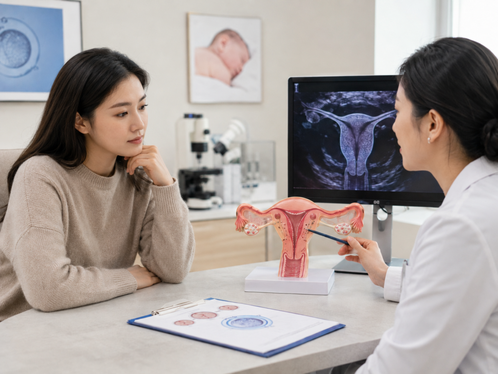
※ Image: AI generated (ChatGPT, OpenAI) / For illustrative reference only.