
KAIST Quantitatively Analyzes Living Eggs and Embryos in 3D Without Damage Objectively Selecting ‘Good Embryos’ Expected to Improve IVF Success Rates
The single most crucial step determining the success or failure of In Vitro Fertilization (IVF) is deciding which embryo to transfer into the uterus. Traditionally, skilled embryologists have relied on microscopes to inspect embryo morphology and cell division states, making selections based on experience. However, an era is opening where the healthiest embryos will be chosen based on numerical data analyzing internal structures rather than just surface appearance.
A research team at KAIST has developed a technology that analyzes the internal structures of living eggs and early-stage embryos in 3D without causing damage, predicting their developmental potential. This is evaluated as a foundational technology that can shift core embryo selection in fertility treatments from human experience to objective data- and AI-driven analysis.
Holotomography: Looking Inside Without Damaging Cells
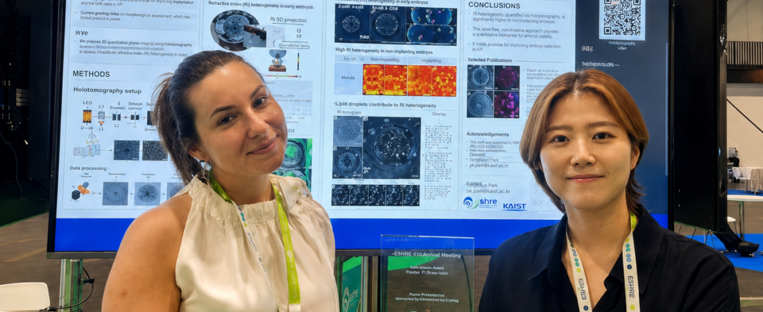
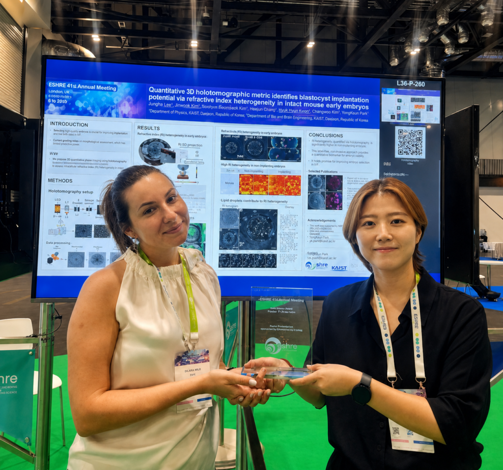
Led by Professor Yong-Keun Park from the Department of Physics at KAIST, the research team recently won the Basic Science Award (Poster Presentation Category) at the 2026 Annual Meeting of the European Society of Human Reproduction and Embryology (ESHRE) held in London, UK. This prestigious award is presented to only one of the most outstanding achievements in the field of basic science presented at the conference.
In fertility treatment, choosing which embryo to transfer among multiple options is a key variable determining pregnancy success rates. However, because eggs and embryos are living cells destined for actual patient transfer, conventional cell analysis methods—such as tissue extraction or fluorescent staining—cannot be used.
Consequently, most fertility clinics currently rely on embryologists to observe embryo size, shape, cell count, and cleavage speed under a microscope before deciding on a transfer candidate. This is also why embryos given the exact same grade can yield vastly different actual implantation outcomes.
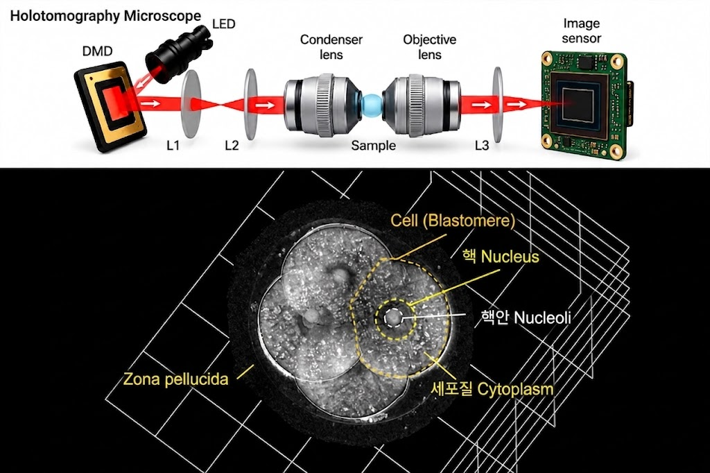
To overcome this limitation, the research team applied an optical technology called ‘Holotomography.’
This technique measures how light refracts as it passes through a cell, reconstructing the cell’s interior in three dimensions. By using no separate stains or chemicals, it precisely analyzes internal structures without harming living cells.
Key Findings: Uniformity and Lipid Droplets
When the research team analyzed mouse early-stage embryos, they found that embryos with lower developmental potential tended to have a more uneven distribution of intracellular materials. Conversely, embryos that successfully developed into blastocysts exhibited relatively uniform internal refractive indices.
Crucially, the team identified that the distribution of ‘lipid droplets’—structures inside cells that store fat—is a major factor driving these refractive index differences. In other words, even embryos that look identical on the outside can have their developmental potential predicted earlier by quantifying their internal structures.
Shifting the Paradigm of Infertility Treatment
This research is significant not merely for developing a novel microscope technology, but for laying the groundwork to evaluate embryos using objective numerical values and AI algorithms rather than subjective human judgment, signaling a potential paradigm shift in infertility treatment.
Once clinical validation using human eggs and embryos is complete, this technology is expected to accurately select embryos with high implantation potential, thereby boosting IVF success rates and reducing the need for unnecessary repeated procedures.
Professor Yong-Keun Park of KAIST stated, “This technology, which quantitatively analyzes the interior of living eggs and embryos in 3D without damage, has received international academic recognition. We are currently conducting validation using human cells and aim to develop a more objective and precise egg and embryo evaluation technology to help improve IVF success rates.”
※ This article was written based on the latest research findings announced by the KAIST research team and the proceedings of the European Society of Human Reproduction and Embryology (ESHRE). It does not replace a specific individual’s diagnosis or treatment, and actual medical judgment must be made through consultation with a specialist.
※ Images: Created using generative AI (ChatGPT, OpenAI); depicts fictional individuals, not real people.