
“Can We Test Oocyte ‘Quality’ Instead of Just Count?”… Clues Uncovered in the Oocyte’s Proteins
- Analyzing oocyte and cumulus cell proteins identifies a ‘proteomic signature’ linked to developmental competence
- Divergences observed in protein synthesis, autophagy, and intracellular vesicular transport pathways
- Researchers suggest potential for developing oocyte quality diagnostics… Primarily preclinical animal models to date, not yet ready for clinical testing
When undergoing fertility care, one of the numbers women hear most frequently is AMH (Anti-Müllerian Hormone). While it offers a valuable estimate of the remaining “quantity” of eggs in the ovaries, it remains silent on the question patients want answered most: “Is the quality of my eggs good?”
Currently, no blood test can quantify egg quality with a numerical score. Clinicians factor in a woman’s age, ovarian reserve biomarkers, and the physical shape and maturity of retrieved eggs. Ultimately, an egg’s true developmental potential is judged only in retrospect—by waiting to see whether it fertilizes properly and develops into a viable blastocyst.
This retrospective limitation is precisely why reproductive scientists have long sought molecular clues inside the egg that could distinguish a truly “good” oocyte from the start. As researchers note, in human assisted reproduction, embryonic development and final clinical pregnancy have remained the de facto gold standard for assessing oocyte quality.
A study published online on August 20 in the international journal Human Reproduction offers a fresh clue. An Australian research team from the University of New South Wales compared the entire protein composition—the proteome—of oocytes with differing developmental capacities, pinpointing a characteristic protein signature directly tied to egg quality.
Even an Egg That ‘Looks Good’ May Be Different Within
In reproductive biology, an egg’s developmental competence means far more than just achieving nuclear maturity. It refers to an oocyte’s internal capacity to sustain early cleavage after fertilization—possessing the essential pool of maternal proteins, messenger RNAs, and metabolic reserves required to drive early life until the embryo’s own genome fully activates.
The dilemma in clinical practice is that observing an oocyte’s exterior morphology under an inverted microscope cannot reliably assess this internal readiness.
To investigate this, the research team established mouse models with known differences in developmental potential: oocytes matured naturally in vivo, oocytes matured in vitro (IVM), and oocytes subjected to CAPA-IVM, a biphasic in vitro maturation system that incorporates a pre-maturation holding phase. Evaluating embryonic development confirmed that in vitro matured oocytes had a diminished capacity to reach the blastocyst stage compared to naturally matured eggs—showing an approximate 40% gap in blastocyst formation rates.
When the researchers looked into their proteomes, however, distinct molecular disparities emerged that would have been entirely invisible under a microscope.
Analyzing ~1,600 Oocyte Proteins: 34 Were Consistently Altered
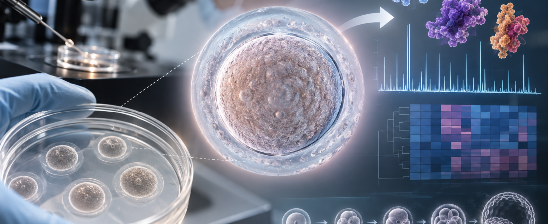
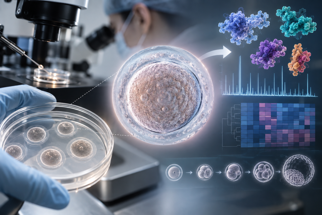
Using high-resolution mass spectrometry, the investigators mapped roughly 1,600 proteins in mouse oocytes and approximately 3,100 proteins in the surrounding cumulus cells. Across these datasets, they identified 34 specific proteins that consistently differed between in vitro matured oocytes and their high-competence, in vivo matured counterparts.
The divergences were especially pronounced in pathways governing translation (protein synthesis), RNA processing, autophagy, and endocytosis—the cellular machinery responsible for internalizing and sorting molecular cargo.
Oocytes matured normally in vivo exhibited relatively higher abundances of proteins central to protein synthesis, such as the translation initiation factor EIF2A and key ribosomal components RPL24 and RPS24. In contrast, in vitro matured oocytes displayed an upregulation of proteins involved in intracellular vesicle trafficking and lysosomal processing.
The authors noted that these shifts likely reflect alterations in how an oocyte synthesizes, maintains, and clears essential proteins as it prepares for fertilization and early embryonic division.
Parallel differences appeared in the surrounding cumulus cells, which nurture the oocyte with metabolic substrates and signaling cues. In cumulus cells supporting in vivo matured eggs, proteins involved in oxidative stress defense and steroid hormone synthesis were relatively enriched. Conversely, cumulus cells from the in vitro group displayed significant proteomic shifts in pathways regulating serine and cholesterol biosynthesis.
In short, the difference between a high-competence egg and one with poor developmental potential does not come down to outer appearance, but rather to the functional integrity of its internal protein systems.
Confirming the Same Markers in Human Oocytes
The investigators next set out to confirm whether the key proteins identified in mouse models were present in human eggs.
Between June and November 2025, they collected 49 immature oocytes donated by 36 women aged 18 to 41 undergoing IVF in Australia—eggs that were clinically unusable for treatment and slated for routine disposal. After maturing subsets of these human oocytes in vitro, the team evaluated target proteins, including EIF2A, RPL24, and CLTB, confirming that these core proteins identified in mouse oocytes are indeed expressed in human oocytes as well.
However, a vital boundary must be drawn here.
This study did not directly compare clinically “good” versus “poor” human eggs to predict live birth outcomes. The human specimens analyzed were not the high-quality, mature eggs used in clinical transfers, but immature gametes that could not be used for patient treatment.
Furthermore, the primary, in-depth proteomic profiling was conducted in animal models. The researchers themselves highlighted as a limitation that they could not yet verify whether the identical molecular alterations are fully conserved across human CAPA-IVM and conventional IVM oocytes.
Not Yet at the Stage of a ‘Single-Drop Blood Test for Egg Quality’
These findings must not be misinterpreted to mean that a simple blood test for egg quality has arrived. The proteins discovered in this research were not circulating biomarkers measured in the bloodstream; they were identified by directly analyzing micro-dissected oocytes and their cumulus cells.
Rather, the true significance of this work lies in opening the door toward evaluating oocyte quality at the molecular level, moving beyond our historical reliance on maternal age, visual morphology, and delayed post-fertilization blastocyst checks.
In particular, if matching diagnostic proteomic signals can be non-invasively detected in shed cumulus cells or spent micro-droplet culture media without disturbing the delicate egg itself, the clinical potential expands substantially. Such technology could eventually lead to objective platforms for identifying which eggs within a retrieved cohort possess the highest biological likelihood of forming a healthy embryo.
The authors concluded that this proteomic signature provides a biological foundation for understanding oocyte competence, offering a roadmap for refining future egg diagnostics and optimizing in vitro maturation protocols. However, transitioning these findings into a clinical test capable of forecasting individual pregnancy and live birth rates will require extensive validation across large cohorts of human mature oocytes alongside tracked clinical outcomes.
For women navigating fertility treatment, knowing “how many good eggs I have” is just as pressing as the raw number of eggs retrieved. Until now, that answer could only be glimpsed days after fertilization had already taken place. This research marks a meaningful step forward by searching for those answers inside the oocyte’s own proteins before development even begins.
Medical Source & Study Reference
- Journal: Human Reproduction (Published online August 20, 2026).
- Study Title: A proteomic signature of oocyte quality from models of varying oocyte developmental competence
- Lead Institutions: School of Biomedical Sciences, University of New South Wales (UNSW Sydney), Australia
- Article ID: deag134 | DOI: 10.1093/humrep/deag134
※ This article was synthesized based on the proteomic research published in the international journal Human Reproduction (August 2026). It does not replace individualized clinical diagnosis or medical care, and specific treatment decisions should always be made in consultation with a qualified reproductive specialist or clinical embryologist.
※ Image: AI generated (ChatGPT, OpenAI) / For illustrative reference only.