
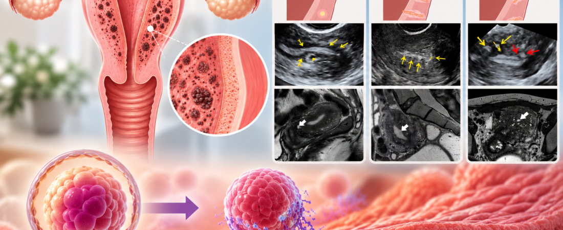
Does the location of adenomyosis matter when you are trying to conceive or undergoing IVF? Two studies reported in July 2026 suggest that disease close to the uterine lining, where an embryo implants, deserves attention alongside its size. Junctional-zone involvement was associated with recurrent implantation failure and pregnancy loss, while diffuse adenomyosis was linked to lower odds of live birth. These findings come from conference abstracts and show associations, not proof of a cause or a treatment benefit.
After a diagnosis of adenomyosis, the first questions are often about size: ‘How many centimeters is it?’ and ‘How severe is it?’ The newer research suggests that asking where it is may be just as useful. Adenomyosis extending close to the implantation site was more often associated with reproductive difficulties, indicating that all cases should not necessarily be viewed in the same way.
What is the junctional zone beneath the uterine lining?
It helps to picture the uterus in layers. The innermost layer is the endometrium, or uterine lining. After an embryo transfer during in vitro fertilization (IVF), this is where implantation takes place.
Outside the endometrium lies the thick muscular wall of the uterus. The inner muscular region at the boundary with the endometrium is called the junctional zone, or JZ. In everyday terms, it is the layer immediately beneath the lining where an embryo implants.
This is more than a dividing line. It participates in the uterus’s small contractions and movements, which are involved in sperm transport and the implantation process.
That raises a question: is adenomyosis in the outer uterine muscle associated with the same reproductive outcomes as disease extending into the region directly beneath the endometrium? One study published in Human Reproduction in July 2026 set out to explore this distinction.
For background on the diagnosis, see our explanation of how adenomyosis differs from uterine fibroids.
What did the study of 280 women find?
Researchers at Policlinico Tor Vergata in Rome, Italy, analyzed 280 women with confirmed adenomyosis. They compared four groups of 70 women each: women with infertility, recurrent implantation failure, recurrent pregnancy loss, and a history of live birth.
The recurrent implantation failure group included only women who had received euploid—chromosomally normal—or donor blastocysts. The purpose was to reduce, to some extent, the possibility that differences in embryo-related factors explained the failed implantation, allowing the researchers to examine differences in the uterus.
Adenomyosis involving the outer uterine muscle was more common in the group with a history of live birth. In contrast, disease involving the junctional zone immediately beneath the endometrium was more common in the recurrent implantation failure and recurrent pregnancy loss groups.
Involvement of the entire junctional zone was associated with recurrent pregnancy loss, with an odds ratio (OR) of 2.35. Severe adenomyosis was also associated with recurrent implantation failure, with an OR of 2.22.
An odds ratio compares the relative odds of an outcome between groups. An OR of 2.35 does not mean that an individual woman’s probability of miscarriage is exactly 2.35 times higher. It is a statistical comparison of odds, not a direct estimate of her personal chance of pregnancy loss.
The label ‘recurrent implantation failure’ also needs careful interpretation. Our related article explains the diagnostic framework for recurrent implantation failure.
What did the analysis of 15,363 women add?
A larger analysis also drew attention to the location and pattern of adenomyosis. A separate 2026 systematic review and meta-analysis combined 25 studies involving 15,363 women, including 5,959 with adenomyosis.
The researchers did not simply divide women into those with and without adenomyosis. They considered features such as focal adenomyosis, concentrated in one area, and diffuse adenomyosis, spread more widely through the uterine muscle. They also examined how close the disease was to the endometrium.
Women with diffuse adenomyosis had lower odds of live birth than women without adenomyosis, with an OR of 0.52. This does not mean that the live birth rate was 52%. It compares the relative odds of live birth in the two groups.
When the researchers considered the studies qualitatively, adenomyosis close to the endometrium also tended to be associated with poorer reproductive outcomes than disease located more peripherally in the uterine muscle.
Why might proximity to the endometrium matter?
Why should a difference in location be associated with a difference in reproductive outcomes? The precise explanation is not fully established.
Adenomyosis can involve changes in uterine muscle structure, inflammation, altered blood flow and abnormal uterine contractions. If these changes occur immediately beside the lining where an embryo needs to implant, they could affect the environment for implantation or the maintenance of early pregnancy.
A similar possibility applies to diffuse adenomyosis: changes extend across a larger area of uterine muscle than in disease confined to one location.
One distinction is essential, however. Observing adenomyosis near the endometrium more often alongside implantation failure or pregnancy loss is not the same as proving that the adenomyosis caused those outcomes. These studies identified associations; they did not establish direct causation.
Look at location and pattern as well as size
The message is straightforward: recording only whether adenomyosis is present, or how many centimeters it measures, may not fully describe the condition.
It may also be useful to ask whether it is focal or diffuse, whether it involves the outer uterine muscle, and whether it extends close to the endometrium where implantation occurs. In other words, consider its ‘address’ alongside its size.
That does not mean that adenomyosis near the endometrium makes pregnancy impossible or means that a miscarriage will occur.
The first study was a retrospective analysis of medical records from a single center involving 280 women. The meta-analysis included 15,363 women, but the studies differed in IVF protocols, embryo-transfer approaches and the criteria used to diagnose adenomyosis. These differences limit how directly the findings can be applied to an individual patient.
Crucially, neither study was a treatment trial showing that treating adenomyosis in a particular location increased implantation or live birth rates. The findings therefore do not justify an immediate conclusion that a lesion must be treated simply because of where it is.
Instead, the questions are becoming more specific: ‘Do I have adenomyosis?’ can be followed by ‘Where is it? How close is it to the uterine lining? Is it concentrated in one area or spread through the uterine muscle?’
The reproductive implications of adenomyosis may not be the same in every case. There is reason to look at where it sits in the uterus as well as how large it is.
Related reading
A separate article examines adenomyosis location and severity in women undergoing euploid embryo transfer.
Research sources and limitations
This article is based on two conference abstracts published on July 8, 2026, in Human Reproduction, volume 41, Supplement 1:
- A. Selntigia and colleagues, including researchers at Policlinico Tor Vergata, Rome, Italy. Adenomyosis and fertility: are there any differences in adenomyosis type and location in patients with distinct reproductive outcomes? Retrospective study of 280 women. DOI: 10.1093/humrep/deag083.325.
- N. B. Brandt and colleagues, including researchers at Odense University Hospital and Copenhagen University Hospital, Denmark. Ultrasonographic features of adenomyosis and pregnancy outcomes in women with infertility or pregnancy loss: A systematic review and meta-analysis. Review of 25 studies involving 15,363 women. DOI: 10.1093/humrep/deag083.876.
Both reports are conference abstracts, not evidence that directly predicts an individual patient’s chance of pregnancy or the effectiveness of a particular treatment.
Image disclosure: The image used in this article was created with generative artificial intelligence (ChatGPT, OpenAI). Any people depicted are fictional and are not real individuals.
Copyright © 2026 Fertility Information Talk Talk — Fertility News | THE FERTILITY NOTES. All rights reserved. Unauthorized republication, reproduction, redistribution, scraping, AI training, or use of any content—including articles, photographs and images—for data purposes is strictly prohibited. Violations may result in legal liability.