
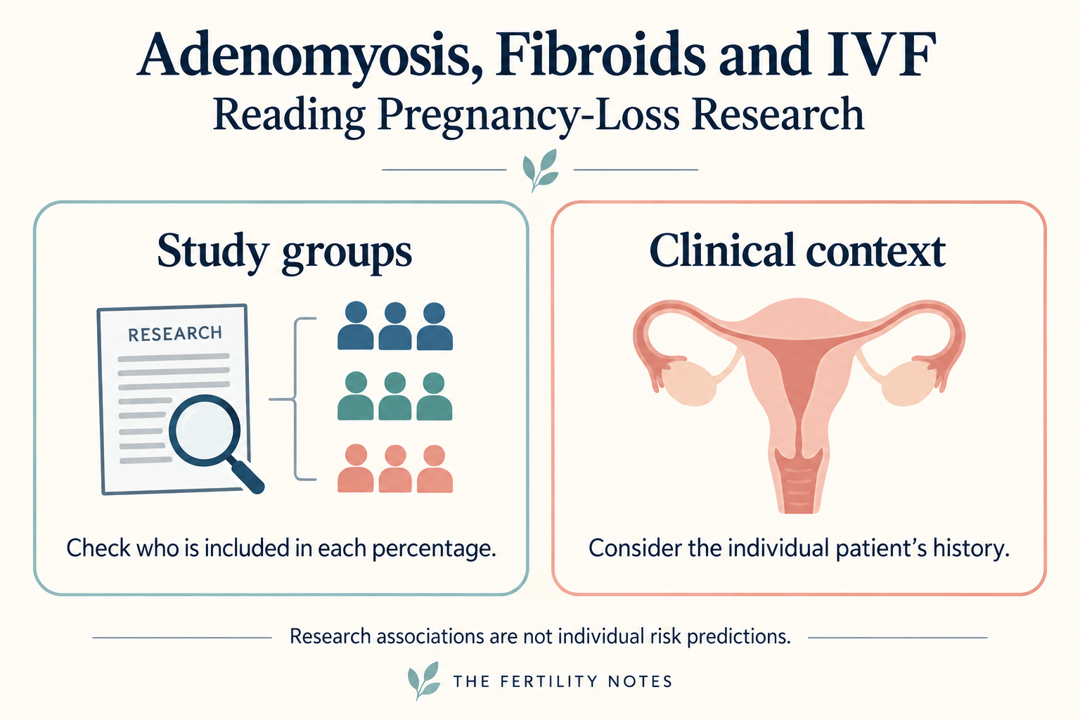
Adenomyosis and fibroids raise different questions in IVF. Understanding pregnancy-loss research starts with checking which patients each percentage describes.
In the field of In Vitro Fertilization (IVF), uterine fibroids are an important clinical variable. Depending on their size, location, symptoms and effect on the uterine cavity, specialists weigh the potential benefits and risks of surgical removal. Myomectomy is not a routine starting point for every patient with a fibroid.
Recent research also draws attention to adenomyosis when considering pregnancy after IVF.
The two diagnoses need to be assessed on their own terms, rather than treating one as universally more important than the other.
What did the IVF pregnancy-loss study actually compare?
Rubin and colleagues studied 971 clinical pregnancies after IVF at one academic center during 2015–2023. Their retrospective cohort compared 14 second-trimester losses with 853 live births; uterine findings used 595 evaluated live-birth controls.
Adenomyosis was present in 5 of 14 loss cases (35.7%) and 38 of 595 evaluated controls (6.4%). These figures describe the frequency of adenomyosis within each outcome group. They are not miscarriage rates among patients with adenomyosis versus patients without it. Primary study.
That distinction matters: reversing the denominator can make an association look like an individual patient’s probability of pregnancy loss.
How do fibroids and adenomyosis differ?
A fibroid is essentially an isolated localized tumor. While it occupies physical space and can mechanically impede embryo implantation depending on its location, it remains a structurally demarcated entity that can be surgically resected.
Adenomyosis differs from a fibroid. It involves endometrial-type glandular tissue within the myometrium, the muscular wall of the uterus. It can be focal or diffuse; describing the whole uterus as hostile or incapable of pregnancy is not an accurate diagnosis. ISUOG patient information.
In clinical practice, distinguishing these conditions can guide the questions asked about fertility and pregnancy.
A fibroid can be measured and mapped on ultrasound, but its diameter alone does not determine whether removal will help. ASRM guidance treats cavity distortion as an important consideration and does not generally advise myomectomy solely to improve pregnancy outcomes in asymptomatic patients with non-cavity-distorting fibroids.
Adenomyosis can also be assessed with ultrasound, sometimes with MRI when the findings are unclear. Imaging may describe its location and extent. These features deserve attention, but they do not establish that the uterus will fail to sustain a pregnancy. ISUOG imaging overview.
Why distinguish implantation from continuing pregnancy?
Embryo quality, transfer timing and uterine factors are all considered in IVF planning. Assessing factors associated with continuing pregnancy is also relevant to this discussion.
The practical point is to consider these questions together. This comparison cannot rank uterine health and embryo quality as competing explanations for every IVF outcome.
Crucially, this cohort examined pregnancy after successful conception. It does not show that a patient with adenomyosis cannot maintain a pregnancy.
Pregnancy loss can bring a profound psychological and physical burden, particularly after fertility treatment. That deserves compassionate care without telling patients that a diagnosis makes loss inevitable or assuming how any one person will experience it.
What can these findings tell patients about treatment?
How, then, can these findings inform a discussion with a fertility specialist?
The discussion can include both whether a fibroid needs treatment and which findings might matter during pregnancy. Useful questions are: Does the lesion affect the uterine cavity? What is known about my individual history? What benefit has been demonstrated for the proposed treatment?
The cohort did not test whether hormonal suppression, endometrial priming, anti-inflammatory treatment or blood-flow interventions improve outcomes. An association cannot establish that these approaches should become routine care.
Reproductive care has to consider the path after embryo transfer as well as the laboratory process. The central concern remains relevant: a pregnancy may begin with successful fertilization, but it is fulfilled only through sustained gestation.
This study does not justify dismissing fibroids or labeling adenomyosis as an inevitable barrier to live birth. With only 14 second-trimester losses, retrospective confounding and unstable estimates remain important limitations. Decisions still require an assessment of the individual patient.
Research sources
Rubin S, Nussbaum O, Noruzi K, et al. F&S Reports 2026;7(1):25–30. Online December 6, 2025. DOI: 10.1016/j.xfre.2025.12.003.
The research team included New York Medical College, Westchester Medical Center and WestMed Reproductive Services. Background information above is linked separately to ASRM guidance and ISUOG resources.
Related reading
- Adenomyosis location, severity and observational IVF outcomes
- Fibroid treatment and five-year fertility outcomes
Editor’s Note: This content is an analytical commentary prepared by a specialized medical journalist through the collection and evaluation of domestic and international reproductive medicine research, clinical policies, and statistical data. All medical diagnoses and treatment plans must exclusively be established through direct consultation with a qualified fertility specialist.
Image Source: AI-generated (ChatGPT, OpenAI) / Provided solely as a supplemental visual aid for conceptual understanding.