
- CHA Medical Center researchers analyze 2,898 single blastocyst transfers at ESHRE 2026
- Whole-chromosome high-level mosaic ongoing pregnancy rate was just 11.1% vs. 51.0% for euploid embryos
- Segmental and low-level mosaic embryos showed no statistically significant differences in reproductive outcomes compared to euploids
- Discard decisions cannot be made based on the word ‘mosaic’ alone… Subtype and percentage must be evaluated together
For patients undergoing preimplantation genetic testing for aneuploidy (PGT-A) during In Vitro Fertilization (IVF), one of the most perplexing and frustrating lab results is a “mosaic embryo.” It is not diagnosed as chromosomally normal, yet it is not classified as fully aneuploid either. It simply means that within the biopsied cells, some had a normal chromosome count while others harbored chromosomal anomalies.
For years, couples receiving a mosaic diagnosis have wrestled with fraught dilemmas: “Is it safe to transfer?”, “How high is the risk of miscarriage or congenital birth defects?”, or “Should we simply discard it and start another retrieval cycle?”
A large-scale analysis recently presented by a South Korean research team reveals that not all mosaic embryos share the same reproductive fate.
Researchers from the CHA Fertility Center Seoul Station at CHA University presented a study at the 2026 Annual Meeting of the European Society of Human Reproduction and Embryology (ESHRE) comparing pregnancy outcomes after transferring single euploid versus single mosaic blastocysts. The findings were featured in the July 2026 ESHRE special abstract issue of Human Reproduction.
The investigative team analyzed 2,898 single frozen blastocyst transfers conducted between August 2020 and June 2025. This cohort comprised 2,569 euploid embryo transfers and 329 mosaic embryo transfers.
The mosaic embryos were categorized into four distinct subtypes based on structural extent and the percentage of abnormal cells:
- Segmental Low-Level Mosaic: Partial chromosome deletion or duplication with a low abnormal cell fraction (154 cycles)
- Segmental High-Level Mosaic: Partial chromosome anomaly with a high abnormal cell fraction (30 cycles)
- Whole-Chromosome Low-Level Mosaic: An entire extra or missing chromosome with a low abnormal cell fraction (118 cycles)
- Whole-Chromosome High-Level Mosaic: An entire extra or missing chromosome with a high abnormal cell fraction (27 cycles)
The Worst Performer: ‘Whole-Chromosome + High-Level’
The clinical divide was stark.
For embryos carrying high-level mosaicism involving an entire chromosome, the clinical pregnancy rate reached only 29.6%—halving the 59.6% seen in the euploid control group.
The disparity widened further when evaluating whether pregnancies progressed beyond the first trimester. While the ongoing pregnancy rate was 51.0% for euploid embryos, it dropped to a mere 11.1% for whole-chromosome high-level mosaic embryos.
Miscarriage rates mirrored this steep decline: while euploid transfers resulted in a 14.4% pregnancy loss rate, whole-chromosome high-level mosaic transfers suffered a miscarriage rate of 62.5%.
This gap remained pronounced even after statistically adjusting for maternal age, paternal age, body mass index (BMI), anti-Mullerian hormone (AMH) levels, and endometrial thickness. The odds of achieving an ongoing pregnancy with a whole-chromosome high-level mosaic embryo were significantly lower than with a normal embryo.
Conversely, embryos categorized as segmental low-level, segmental high-level, or whole-chromosome low-level mosaic showed no statistically significant differences in clinical pregnancy or ongoing pregnancy rates compared to fully euploid embryos.
This demonstrates that a blanket label of “mosaic” is clinically insufficient to predict whether an embryo can result in a healthy pregnancy.
‘Chromosome 9 -20%’ Does Not Mean What You Think
If a PGT-A lab report indicates a result like “partial deletion of chromosome 9, 20% mosaic,” patients should not immediately assume the embryo is unviable.
Clinicians must evaluate whether the abnormality involves an entire chromosome or only a regional segmental fragment, determine the exact percentage of mosaicism detected, and check whether other fully euploid embryos are available in the cohort.
A borderline low-level segmental mosaicism (such as 20%) behaves biologically very differently from a high-level mosaic involving a complete chromosome. However, because different genetic reference laboratories use varying baseline thresholds to define mosaicism and categorize “low” versus “high” levels, patients cannot automatically assume that a 20% reading is completely without risk.
Furthermore, PGT-A does not test every cell in the embryo.
The biopsy removes only 5 to 10 cells from the outer trophectoderm layer, which eventually forms the placenta, leaving the inner cell mass that develops into the fetus untouched. Consequently, a trophectoderm biopsy provides a representative sample rather than an absolute, 100% guarantee of the chromosomal architecture of the entire developing baby.
Reflecting these nuances, ESHRE guidelines recommend comprehensive genetic counseling before scheduling a mosaic embryo transfer to explain the biological limitations of trophectoderm sampling, the significance of the specific chromosome involved, and the distinction between whole-chromosome and segmental variations.
Neither Automatic Discard Nor Careless Transfer
The core message of this study is straightforward: embryos bearing the “mosaic” label should not all be treated as equal.
When a patient has confirmed euploid embryos in storage, those euploid blastocysts naturally take priority for transfer. However, for poor responders or patients who have exhausted their euploid options and hold only mosaic embryos, examining the fine print of the mosaic classification can reveal viable opportunities for pregnancy.
At the same time, this study was a retrospective observational cohort rather than a randomized controlled trial, and the data were presented in conference abstract format at ESHRE 2026. The authors cautioned that their conclusions are intended to help prioritize embryos in clinical practice rather than guarantee definitive outcomes.
Receiving a mosaic PGT-A result should not lead to an immediate discard order, nor should it lead to an unconsidered transfer. The safest path forward is a structured discussion with a reproductive endocrinologist and genetic counselor to review whether the anomaly is partial or complete, determine the exact percentage of affected cells, identify the specific chromosome involved, and weigh the clinical options based on the patient’s entire reproductive journey.
※ This article was synthesized based on the research abstract presented by researchers from CHA Fertility Center Seoul Station, CHA University School of Medicine, at the 2026 Annual Meeting of the European Society of Human Reproduction and Embryology (ESHRE) and published in the July 2026 conference special issue of Human Reproduction. It does not replace individualized clinical diagnosis or medical care, and specific treatment decisions should always be made in consultation with a qualified reproductive specialist or clinical geneticist.
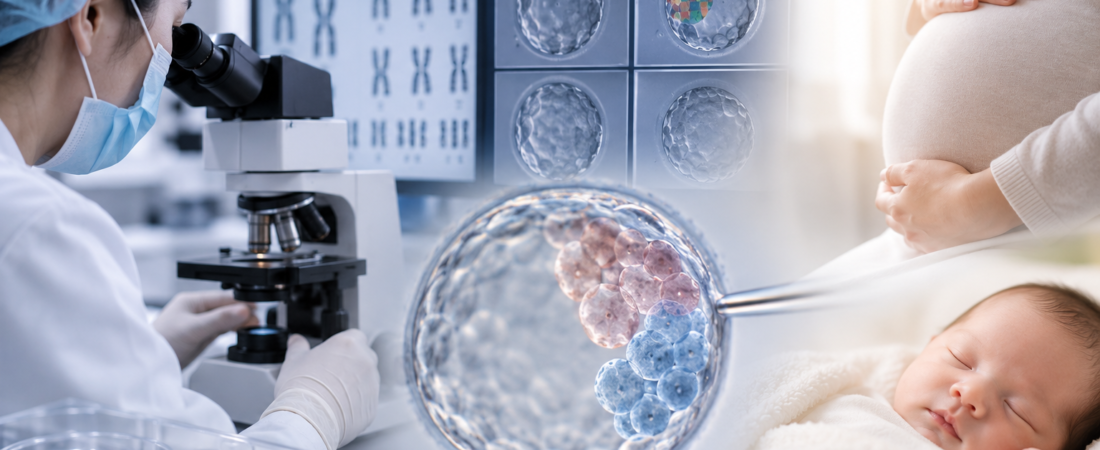
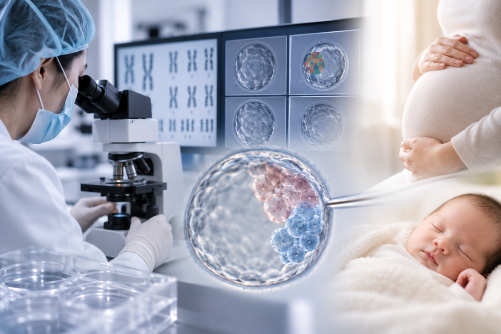
※ Image: AI generated (ChatGPT, OpenAI) / For illustrative reference only.