
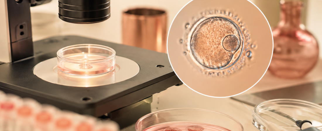

Can a 1PN embryo—an embryo observed with one pronucleus after fertilization—lead to a live birth? A retrospective IVF study examined 13,203 1PN embryos and reported outcomes for selected blastocysts after genetic assessment. Results differed between conventional IVF and ICSI, and findings from tested, transferred blastocysts should not be generalized to every 1PN embryo.
- Challenging the Dogma of 1PN Discard: Multicenter Australian study of 13,203 1PN embryos across 10,730 cycles found biparental origin in most tested conventional IVF 1PN blastocysts; biparental origin does not itself establish euploidy
- Conventional IVF vs. ICSI Divergence: 96.9% of tested conventional IVF 1PN blastocysts possessed genetic material from both parents (only 3.1% uniparental), whereas ICSI 1PN blastocysts had a 34.1% uniparental rate
- Outcomes After Selected Transfers: The study did not detect adjusted live-birth or reported perinatal differences in selected euploid, biparental 1PN transfers; this does not establish equivalence or long-term safety
- When No Usable 2PN Embryos Remain: Approximately 1 in 6 cycles (15.6% to 16.7%) with 1PN embryos had no usable 2PN embryos; this does not establish a separate treatment benefit for low responders
In standard In Vitro Fertilization (IVF) laboratory protocol, checking for fertilization 16 to 18 hours post-insemination represents a critical milestone. The visualization of two pronuclei (2PN)—one containing maternal chromosomes and the other paternal chromosomes—is a standard morphological sign used to assess fertilization; this observation alone does not prove euploidy.
Conversely, zygotes displaying only one pronucleus (1PN) have historically been viewed with clinical suspicion. Because a single pronucleus can theoretically indicate abnormal parthenogenesis (spontaneous activation of the egg without sperm genome integration) or gynogenetic/androgenetic haploidy, many embryology laboratories have routinely discarded 1PN embryos or excluded them from clinical transfer.
A landmark multicenter study published in the August 2026 issue of Human Reproduction challenges this routine discard policy. Analyzing 13,203 1PN embryos across 10,730 clinical cycles from Australian fertility networks (Genea and the University of Adelaide), researchers assessed genetic findings in selected blastocysts and live births after selected transfers. Parental-origin and aneuploidy results differed between IVF and ICSI; the findings do not establish normality or safety for all 1PN embryos.
Why an IVF fertilization check may show one pronucleus
A 1PN observation under the microscope at a fixed time point does not automatically mean the embryo is biologically abnormal. Several physiological mechanisms can create the visual appearance of a single pronucleus:
- Asynchronous Pronuclear Formation: Maternal and paternal pronuclei form at slightly different times; an early or late check may catch only one visible pronucleus.
- Early Pronuclear Apposition or Fusion: The two pronuclei may migrate together and fuse earlier than standard observation windows, appearing as a single larger nuclear structure.
- Premature Pronuclear Envelope Breakdown: One pronuclear membrane may break down prematurely prior to syngamy.
A single static visual check at hour 17 captures a momentary snapshot that may misclassify a normally fertilized zygote.
1PN blastocysts after conventional IVF versus ICSI
To determine the true genetic origin of 1PN embryos, the researchers performed Preimplantation Genetic Testing for Aneuploidy (PGT-A) combined with Short Tandem Repeat (STR) genotyping to trace parental origin:
| Parameter | Conventional IVF 1PN Blastocysts | ICSI 1PN Blastocysts | Standard 2PN Blastocysts (Reference) |
| Biparental Genomic Origin | 96.9% (440 / 454) | 65.9% (108 / 164) | Not reported for 2PN biparental origin |
| Uniparental Inheritance | 3.1% (14 / 454) | 34.1% (56 / 164) | Not reported for 2PN uniparental inheritance |
| Aneuploidy Rate | 37.3% | 45.5% as reported (source discrepancy; see note below) | 31.5% to 33.9% |
| Ploidy Difference vs. 2PN | Not statistically significant (after age adjustment) | Significantly higher aneuploidy | Baseline reference |
Source discrepancy: The paper reports 45.5% for ICSI 1PN aneuploidy, but its Table 2 lists 122/246 beside that percentage; the fraction calculates to approximately 49.6%. We have not verified which value needs correction. The table above retains the reported percentage with this warning, rather than presenting a recalculation as a corrected study result.
- Conventional IVF (Insemination): Among 1PN embryos that reached the blastocyst stage and underwent genetic tracing, 96.9% were confirmed to carry both maternal and paternal DNA. The aneuploidy rate (37.3%) did not differ significantly from standard 2PN sibling embryos (33.9%) after controlling for maternal age.
- Intracytoplasmic Sperm Injection (ICSI): 1PN embryos derived from ICSI carried a substantially higher risk of uniparental inheritance (34.1%) and elevated aneuploidy (reported as 45.5%; see the source-discrepancy note above). The authors discuss premature oocyte activation and sperm-decondensation failure as possible explanations; this cohort does not establish the cause of each 1PN finding.
How often did 1PN embryos reach the blastocyst stage?
The study reported lower Day 5 blastocyst development for 1PN than for 2PN embryos:
- Blastocyst Development Rate (Day 5):
- Conventional IVF: 19.3% for 1PN vs. 63.3% for 2PN
- ICSI: 9.7% for 1PN vs. 60.6% for 2PN
Reaching the blastocyst stage does not itself establish euploidy or biparental origin: genetic abnormalities remained among the tested blastocysts.
Live birth and reported perinatal outcomes after selected 1PN transfers
For the subset of 1PN blastocysts that were confirmed to be euploid and biparental, the study reported the following outcomes compared with standard 2PN transfers:
- Live Birth Rate per Transfer:
- Conventional IVF 1PN: 40.9% vs. 50.4% for 2PN (the difference was not statistically significant after adjustment for maternal age, embryo freeze day and patient ID; this does not prove equivalence).
- ICSI 1PN: 40.7% (based on 54 validated transfers).
- Perinatal and Neonatal Health: Available birth-outcome data covered 81 IVF 1PN and 22 ICSI 1PN births. Reported gestational age, term-delivery rate, birth weight and small/large-for-gestational-age outcomes did not differ significantly from 2PN controls; long-term child health was not established.
- Complication Rates: Reported maternal/neonatal complication rates were 9.9% in IVF 1PN and 9.6% in IVF 2PN births, without a detected difference. No hydatidiform mole (molar pregnancy) was reported among 257 1PN transfers; this does not exclude rare risks.
Why parental-origin testing matters alongside PGT-A
The study highlights that copy-number-only PGT-A does not establish parental origin or exclude every ploidy error:
- The Copy Number Blind Spot: Copy-number PGT-A assesses relative chromosome dosage; it does not directly verify a complete set of 46 chromosomes. However, it cannot distinguish whether a diploid set of 46 chromosomes originated biparentally (23 maternal + 23 paternal) or uniparentally (e.g., 46 maternal chromosomes via endoreduplication, or 46 paternal chromosomes).
- Molar Pregnancy and Imprinting Risks: Androgenetic diploid embryos (paternal only) can lead to complete hydatidiform moles, and parental-origin concerns are distinct from the chromosome-dosage result.
- Testing in This Study: The clinic used PGT-A plus STR parental-origin analysis before 1PN transfer. Its STR classification had not been independently validated against another technology for this application; testing choices require discussion with the clinical and genetics teams.
1PN embryos when no usable 2PN embryos remain
The most urgent clinical finding for patients is cycle utilization:
- In 15.6% of conventional IVF cycles and 16.7% of ICSI cycles where a 1PN embryo was observed, no usable 2PN embryos were available.
Where no usable 2PN embryo remains, a 1PN embryo may be the only potential option from that cycle. This study did not establish a separate treatment rule for POSEIDON groups or patients retrieving only one or two oocytes.
The study describes an approach for individualized consideration, rather than an immediate universal change in practice:
- Extended Blastocyst Culture: Allowing 1PN zygotes to grow to Day 5/6 while recognizing that blastocyst survival does not prove genetic normality.
- Trophectoderm Biopsy with Parental-Origin Testing: Performing PGT-A combined with STR/parental origin testing for surviving blastocysts.
- Individualized Transfer Consideration: Discussing a tested biparental, euploid 1PN blastocyst as a potential option, particularly when no usable 2PN embryos remain.
ESHRE laboratory guidance discussed in this article
The European Society of Human Reproduction and Embryology (ESHRE) published updated laboratory recommendations in the August 2026 issue of Human Reproduction.
ESHRE supports cautious consideration of 1PN embryos after blastocyst culture, with PGT-A suitable for assessing biparental diploidy if available. Use should be discussed with the clinical team and patient, governed by an internally approved policy and fully traceable. This is not a guarantee of safety or a universal testing mandate.
For related questions about interpreting embryo assessments, read mosaic embryo transfer and PGT-A result interpretation and PGT-A outcomes per transfer versus per egg retrieval.
Medical Source & Study Information
- Journal: Human Reproduction (Official Journal of the European Society of Human Reproduction and Embryology, ESHRE), Vol. 41, Issue 8, pp. 1293–1309 (August 2026).
- Study Title: A large retrospective study on 1PN embryo transfer supports the need for updated harmonized best practice guidelines
- Lead Institutions: Genea Fertility and The University of Adelaide, Australia
- DOI: 10.1093/humrep/deag098. Laboratory guidance: ESHRE recommendations on Good Practice in the IVF laboratory, doi:10.1093/humrep/deag096.
Study limitations: This retrospective analysis used records from multiple Genea clinics within one organization and examined selected tested and transferred blastocysts. Selection bias, small transfer subgroups and incomplete birth follow-up limit interpretation. STR parental-origin classification was not independently validated for this application, and long-term child health was not established. The authors reported no specific funding for the work and no conflicts of interest.
※ This article was synthesized based on the multicenter cohort study published in Human Reproduction (August 2026) and updated laboratory consensus guidelines from ESHRE. It does not replace individualized clinical diagnosis or medical care, and specific embryological decisions should always be made in consultation with a qualified reproductive endocrinologist and clinical embryologist.
※ The images associated with this article were generated using generative AI (ChatGPT, OpenAI) as illustrative visual references and do not depict real individuals.