
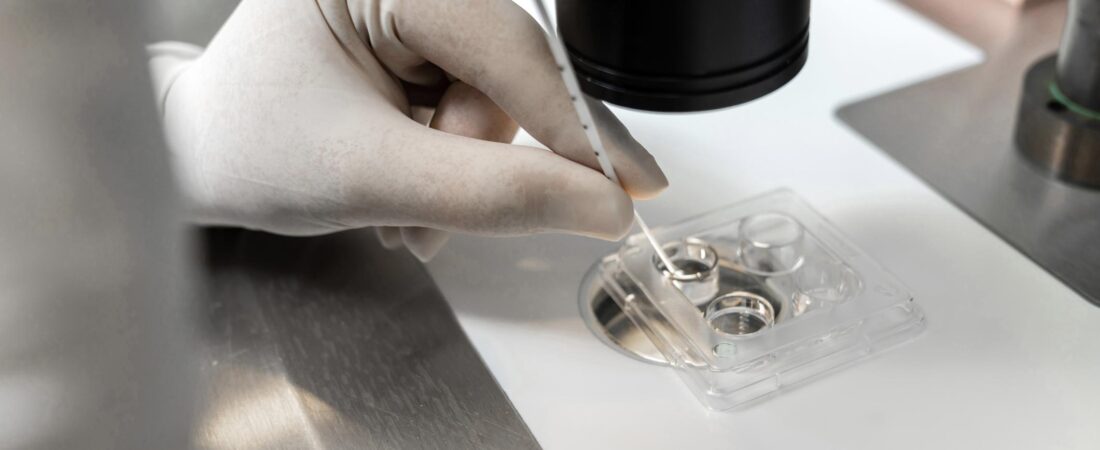
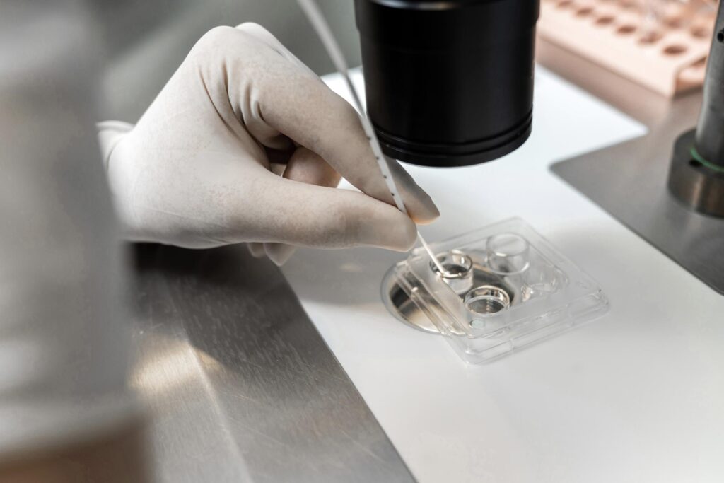
There is a moment during an In Vitro Fertilization (IVF) cycle that is profoundly quiet. It is the mere span of a few seconds when a single sperm is micro-injected into an oocyte. On the laboratory monitor, it appears as though absolutely nothing unusual is taking place.
The oocyte retains its perfectly spherical shape, and the official chart subsequently records the fertilization as normal. At that specific junction, no one questions the validity of the process. Yet, it is precisely during those few seconds that the ultimate outcome of the cycle has already been determined.
Intracytoplasmic Sperm Injection (ICSI) is not merely an automated laboratory protocol; it is a highly specialized art executed by human hands. To be more scientifically precise, it is governed by longitudinal tactile intuition.
In hands that lack this refined intuition, reproductive failure initiates silently. The defining challenge of this failure mode is that it remains completely invisible. It leaves no trace on the patient’s medical charts, nor can it be captured by standard statistical metrics. Consequently, this hidden variable operates in a manner that is deeply elusive and, for the patient, incredibly cruel.
In unrefined hands, the injection needle does not seamlessly glide into the cell; rather, it forcefully punctures the structure. The operator drives the needle forward using blunt mechanical force instead of calculating the optimal fluid entry angle.
At that precise micro-second, a subtle structural fracture occurs within the oocyte’s internal matrix. On the surface, the cellular oolemma snaps shut as if entirely undisturbed. The external appearance looks flawless.
Fertilization is achieved, and early cellular cleavage even begins. This superficial stability makes the underlying damage exceptionally easy to overlook.
However, the biological timeline of this resulting embryo is inherently compromised. As the first 24 to 48 hours pass, the precise developmental rhythm begins to fracture. The rate of cell division slows down, the architectural symmetry of the blastomeres blurs, and the embryo ultimately undergoes a complete developmental arrest. While the clinical failure only becomes visible days later, the trajectory toward that failure was established long before.
Advanced micro-injection is not a primitive piercing technique. It is a highly delicate manipulation of a fluid-filled biological structure. A masterfully trained hand enters the cell by evenly dissipating the physical displacement pressure, minimizing any disruptive shearing forces within the ooplasm.
Conversely, when an operator lacks sufficient experience, the internal architecture of the oocyte undergoes a violent hydrodynamic shock at the exact moment of fluid injection. It is structurally equivalent to abruptly striking a glass filled to the brim with water. Externally, the fluid volume appears unchanged, yet the internal equilibrium has been completely shattered.
The downstream consequences of an inexperienced embryologist’s manipulation surface a few days later. The embryo forms, but it lacks the metabolic capacity to sustain development to the blastocyst stage. Because the precise etiology cannot be documented through standard means, the clinical responsibility is easily and frequently deflected away from the laboratory.
The selection of the paternal gamete is similarly a task that cannot be accomplished through superficial visual observation alone. Under a standard high-magnification microscope, almost every motile sperm cell appears viable. They move, they swim, and they are functionally alive.
Yet, their internal genomic integrity operates on an entirely different plane. Certain sperm heads house structurally fragmented paternal DNA, while others are severely compromised by oxidative stress.
A master embryologist looks deeply at the kinetic quality of the motion—meticulously decoding the velocity, the tail-bouncing trajectory, and the most minute flagellar oscillations. In stark contrast, an inexperienced operator simply selects any cell that is moving.
The cascading outcome of this choice is straightforward: the sperm has successfully entered the egg, and initial fertilization has been logged, yet the resulting embryo fails to thrive. In the absence of deeper insights, the clinical narrative naturally shifts the blame entirely toward the quality of the maternal oocyte.
The variable of timing is even more elusive. The exact moment an oocyte is retrieved from the follicle, its biological state enters a phase of rapid, time-sensitive degradation. A variance of mere minutes can fundamentally alter its cellular receptivity. A highly synchronized embryology team interprets this window not as a rigid numerical manual, but as a fluid biological continuum. They possess the clinical intuition to feel exactly when the oocyte is primed for injection.
To an inexperienced operator, however, time is treated as a flat, corporate protocol. They operate under the assumption that as long as the steps are executed within the generalized guidelines, the outcome will remain stable. Yet, living biological systems do not conform to standardized instruction manuals. These microscopic discrepancies eventually compound into massive divergences in clinical success rates.
The core frustration lies in the fact that none of these real-time variables are ever officially recorded.
The medical charts simply state that normal fertilization occurred. The subsequent lab reports merely document delayed embryonic development. The fractions of a second, the minor millimeters of needle deviation, and the microscopic grams of excess pressure applied during the procedure disappear entirely into a clinical void.
Consequently, the patient is left to digest only the final, negative outcome. They are told that the oocytes were of poor quality, and they are forced to accept the absolute conclusion that maternal age was the sole barrier. In reality, the true trajectory of that embryo may have been altered at the very inception of the laboratory cycle.
Advanced micro-injection is never a battle of equipment infrastructure. Utilizing the exact same micromanipulator, the exact same injection pipettes, and the exact same cleanroom incubator space, entirely disparate live birth rates are achieved. The single, overriding variable is the individual executing the procedure.
It is not an operator who has merely repeated the same motion a high number of times who secures success, but a specialist who has deeply analyzed past failures and refined their approach accordingly. True mastery in the embryology lab is not a skill of mindless repetition; it is the physical memory of a highly calibrated hand. And when a laboratory lacks that refined memory, a developing life quietly loses its direction without making a sound.
📚 Medical References
- Palermo G, Joris H, Devroey P, Van Steirteghem AC.
- “Pregnancies after intracytoplasmic injection of single spermatozoon into an oocyte.”
- Lancet. 1992;340(8810):17–18.
- Significance: The very first clinical report of ICSI. This landmark paper marks the origin of the procedure, demonstrating that the entire technique is fundamentally based on precise “manual manipulation.”
- Rienzi L, Vajta G, Ubaldi F.
- “Predictive value of oocyte morphology in human IVF: a systematic review of the literature.”
- Human Reproduction Update. 2011;17(1):34–45.
- Significance: Provides clear clinical evidence establishing that micro-trauma and subtle cytoplasmic changes within the oocyte directly impair subsequent embryonic development.
- Ebner T, Moser M, Tews G.
- “Possible errors in intracytoplasmic sperm injection.”
- Human Reproduction. 2001;16(2):255–262.
- Significance: A core paper that directly addresses technical errors that can occur during the ICSI process, such as injection placement site and mechanical damage to the cytoplasm.
Editor’s Note: This commentary has been comprehensively reconstructed based on publicly available reproductive medicine literature, peer-reviewed international journals, and clinical research data. All medical diagnoses and treatment plans must exclusively be established through direct consultation with a qualified fertility specialist.
Image Source: AI-generated (ChatGPT, OpenAI) / Provided solely as a supplemental visual aid for conceptual understanding.