
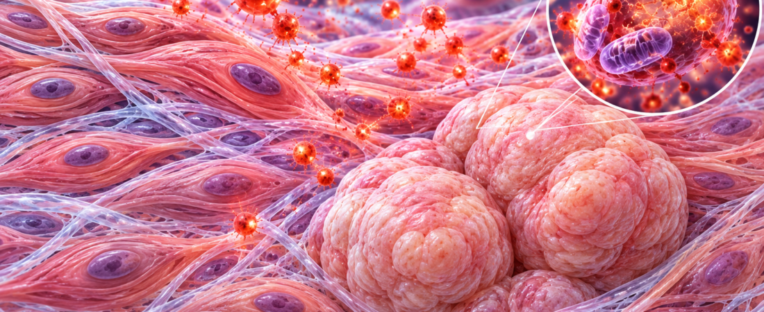
Chronic oxidative stress may contribute to the cellular changes involved in uterine fibroid development. New laboratory research found that weakening antioxidant defenses increased DNA damage and changes in genes associated with fibroids.
Researchers at Northwestern University Feinberg School of Medicine in the United States reported the findings in Scientific Reports on October 16, 2026.
The study looks beyond the established role of female sex hormones in fibroid growth to investigate early changes that may help normal uterine muscle cells acquire tumor-related characteristics.
Oxidative stress also appears in normal tissue surrounding fibroids
Uterine fibroids are benign tumors arising from smooth muscle cells in the uterine wall. They are common during the reproductive years and, depending on their size and location, can cause heavy menstrual bleeding, painful periods, pelvic pain, anemia and infertility.
Estrogen, progesterone and genetic factors are known to contribute to fibroid development. How normal uterine muscle cells become fibroid cells is not yet fully understood.
The researchers focused on oxidative stress: an imbalance between reactive oxygen species generated within cells and the antioxidant defenses that remove them.
Reactive oxygen species also arise during normal cell activity. When they accumulate excessively, they can damage DNA, proteins and other cellular components and alter cell function.
Earlier research observed increased oxidative stress not only in fibroids but also in surrounding uterine muscle tissue. The team considered whether this might contribute to fibroid formation, rather than only result from it.
Reduced antioxidant defenses increased DNA damage
The researchers used human myometrial cells—muscle cells from the uterine wall—to study the effects of chronic oxidative stress.
They reduced the function of manganese superoxide dismutase, or MnSOD, an antioxidant enzyme that helps remove reactive oxygen species, creating a sustained oxidative environment.
They then examined cell growth and gene expression using three-dimensional cell culture and xenografts in immunodeficient mice. In some experiments, cells were followed under prolonged oxidative stress for up to 10 months.
Cells with reduced antioxidant enzyme function accumulated more reactive oxygen species and 8-OHdG, a marker of oxidative DNA damage.
Gene expression analysis also revealed changes in pathways involved in collagen and extracellular matrix formation, cellular senescence, and cell growth and survival.
Changes related to extracellular matrix accumulation were particularly notable because this is a major feature of fibroids. The extracellular matrix surrounds cells and supports tissue structure; fibroids characteristically accumulate excessive amounts of collagen and other matrix components.
The researchers interpreted these findings as evidence of a relationship between chronic oxidative stress and cellular characteristics associated with fibroids.
More mutations in the fibroid-associated MED12 gene
Another important finding concerned MED12, a gene strongly associated with fibroid development.
MED12 helps regulate gene expression, and mutations in this gene occur in a substantial proportion of fibroids.
Mutations were more frequent in experiments involving transplanted uterine muscle cells exposed to prolonged oxidative stress.
Cell proliferation increased modestly, while cell death did not rise substantially. This suggests that some stressed cells may survive in the altered environment while developing molecular features associated with fibroids.
However, the number of MED12 mutations detected was limited. They were not consistently concentrated at the specific mutation sites commonly found in actual fibroids.
The findings therefore do not establish that oxidative stress directly causes fibroids in the human uterus.
A research direction, not proof that antioxidant supplements prevent fibroids
The study suggests that fibroid development should be investigated not only in terms of hormones and genetics but also in relation to the oxidative environment experienced by uterine muscle cells over time.
The finding that impaired antioxidant defenses can affect DNA damage, gene expression and extracellular matrix formation may provide clues to the mechanisms underlying fibroid development.
The researchers suggested that mechanisms regulating the balance between oxidation and reduction within cells could potentially inform future strategies to prevent fibroid development or limit progression.
But this was basic research using cells and animal models. It did not demonstrate that taking antioxidants or eating particular foods prevents fibroids or reduces their size.
Further research is needed to establish how much chronic oxidative stress contributes to fibroid development in people.
The work points toward studying fibroids as a condition involving interacting oxidative and genetic changes in uterine muscle cells, alongside the established influence of hormones on tumor growth.
Source: Jian-Jun Wei, Matthew John Schipma, J. Julie Kim and colleagues at Northwestern University Feinberg School of Medicine. “Human myometrial cell fate under chronic oxidative stress for leiomyomagenesis.” Scientific Reports, October 16, 2026. DOI: 10.1038/s41598-026-74367-w.
Related reading
Read more about research on menstrual symptoms in endometriosis and adenomyosis, two distinct conditions.
The image was created using generative AI (ChatGPT, OpenAI); any people depicted are fictional.
Copyright © 2026 Fertility Information TokTok | THE FERTILITY NOTES. All rights reserved. Unauthorized republication, reproduction, redistribution, scraping, AI training and data use of any content, including articles, photographs and images, are strictly prohibited. Violations may result in legal liability.