
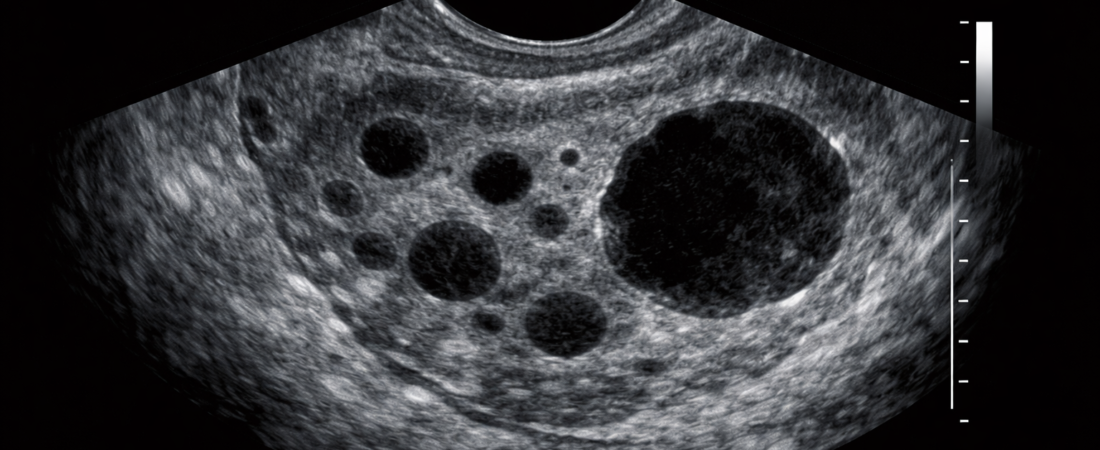
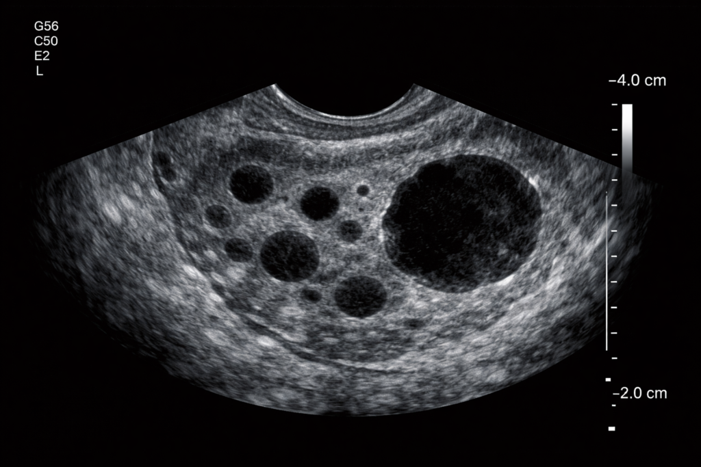
There is a singular, paramount window during a fertility consultation cycle: the transvaginal ultrasound evaluation.
For a reproductive endocrinology specialist, those brief seconds when developing follicles and the endometrial lining materialize on the laboratory monitor determine the entire trajectory of the cycle. Should the egg retrieval be accelerated, or should it be delayed? Is a fresh embryo transfer clinically viable, or must the protocol be aborted entirely? Ultimately, every critical decision initiates within this brief visual window.
Reproductive medicine is fundamentally a discipline of timing. A variance of mere hours can completely invert the final outcome. A clinician must accurately read the subtle micro-structural shifts immediately preceding spontaneous ovulation and decode the incredibly narrow receptive window of the endometrium. If this definitive clinical judgment is forced to rely strictly on a “captured static image” or a “summarized numerical report,” to what degree can the actual precision of the cycle be guaranteed?
Within advanced fertility care, the specific individual executing the ultrasound evaluation carries immense clinical weight. The scan can be performed directly by the primary fertility specialist, or it can be outsourced to a general sonographer or radiologist.
To be fair, separating ultrasound operations into a standalone diagnostic department possesses clear structural advantages. It accelerates patient throughput, drastically minimizes waiting times, secures a standardized quality of imaging records, and empowers a clinic to manage a substantially higher volume of cases. From a purely administrative standpoint, this model represents an entirely rational, efficient choice.
However, the delicate biological calculus of reproductive medicine consistently deviates from this logic of corporate efficiency.
An ultrasound is not a static photograph; it is a live broadcast of moving physiology. Two distinct follicles can measure an identical 18 mm on a report, yet one may already be over-matured and degenerating while the other is precisely primed for retrieval. Similarly, evaluating the endometrium requires a deep reading of tissue responsiveness rather than a flat measurement of raw thickness. Whether the tissue is dynamically perfused with blood and biochemically prepared to capture an embryo is a microscopic nuance that leaves zero trace on a standardized numerical chart. The core problem is that these fluid variables cannot be captured in a post-examination written report—and it is precisely within this unrecorded gap that success and failure diverge.
Consider the case of Patient A (37), who was preparing for her third consecutive IVF cycle. Her outsourced diagnostic chart documented: “7 follicles visualized, average diameter 18 mm.” Based strictly on these optimal numbers, there was zero reason to alter the timeline. The retrieval proceeded exactly as scheduled. The clinical outcome, however, was devastating: out of the 7 tracked follicles, only 3 oocytes were successfully retrieved, yielding a single normal fertilization.
The underlying explanation was simple: beneath that uniform numerical average co-existed a highly mismatched mix of post-mature, post-ovulatory follicles and completely under-developed ones. This profound physiological asymmetry remains entirely invisible on a flat data sheet. It is information that can only be successfully decoded by the specific eyes directly operating the probe in real time. Human biology never strictly conforms to standardized textbook rules—a baseline reality that is exceptionally volatile in patients navigating diminished ovarian reserve.
Would the strategic choice have changed if the primary fertility specialist had been directly evaluating that ultrasound screen? In all clinical probability, yes. They might have aggressively accelerated the retrieval timeline, recalibrated the immediate medication strategy, or at the very least, made the definitive choice that “now is not the time.” This demonstrates that entirely disparate clinical conclusions can be extracted from the exact same biological profile, depending entirely upon who is interpreting the live image.
Advanced fertility care thrives or fractures within the hidden gap between information transmitted as raw data versus insights forged through direct, real-time clinical observation. That microscopic gap ultimately dictates the difference of a single viable embryo—and the success of an entire pregnancy attempt.
Modern medicine is systematically shifting toward automated efficiency, rendering the division of labor an institutional inevitability. Yet, the field of assisted reproduction stands as a fierce exception to this rule. This discipline remains, at its core, a practice of “direct observation and immediate execution.” Consequently, within an IVF cycle, a patient secures a vastly superior clinical advantage when their primary physician personally conducts and interprets every ultrasound scan.
This direct approach becomes a non-negotiable priority for couples navigating recurrent implantation failure (RIF), patients exhibiting highly erratic and unpredictable ovarian responses, advanced maternal age cohorts where the genomic integrity of every single oocyte is critical, or individuals managing localized pelvic pathologies. In these high-stakes scenarios, the capacity for immediate, expert interpretation functions as the definitive variable that reconfigures the final outcome.
📚 Medical References
- Zini A, Sigman M.
- “Clinical utility of sperm DNA damage tests: a comprehensive review.”
- Human Reproduction.
- Significance: Analyzes how advanced paternal variables corrupt early embryonic cleavage, reinforcing the necessity for precise, real-time maternal cycle adjustments to maximize limited embryo utility.
- Paulson RJ, et al.
- “Clinical relevance of oocyte maturation and follicular dynamics in assisted reproductive technology.”
- Fertility and Sterility.
- Significance: Outlines the profound physiological asynchronous development that occurs within a single follicular cohort, demonstrating why raw numerical measurements fail to reflect actual oocyte maturity.
- Gardner DK, Schoolcraft WB.
- “Morphological criteria for embryo selection and the correlation with clinical live birth outcomes.”
- Human Reproduction Update.
- Significance: Explains the critical need for absolute synchronization between the structural quality of the embryo and the real-time receptive state of the endometrium.
- Loverro G, et al.
- “The relationship between follicle size, follicular fluid metrics, and oocyte developmental competence.”
- Journal of Assisted Reproduction and Genetics.
- Significance: Establishes that follicle diameter alone is a highly unreliable isolated predictor of oocyte viability, validating the requirement for comprehensive, direct sonographic evaluation by the treating physician.
Editor’s Note: This content is an analytical commentary prepared by a specialized fertility journalist through the collection and evaluation of domestic and international reproductive medicine research, clinical policies, and statistical data. All medical diagnoses and treatment decisions must exclusively be established through direct consultation with a qualified medical professional.
Image Source: AI-generated (ChatGPT, OpenAI) / Provided solely as a supplemental visual aid for conceptual understanding.