
For a long time, men held onto a single, comforting belief: while a woman’s fertility is heavily bound by age, men remain relatively free from the ticking biological clock. Because functional sperm cells are manufactured continuously throughout a man’s life, this premise sounded remarkably plausible.
However, contemporary breakthroughs in reproductive medicine are quietly dismantling this long-standing assumption. Sperm cells do not escape the aging process; rather, they age through a completely different biological mechanism.
Looking strictly at standard, visible semen parameters, the structural shifts do not appear drastic. Sperm concentration often remains relatively stable, subject to individual variance, and the baseline morphology observed under a standard microscope does not show a sudden, catastrophic collapse.
Yet, this is precisely where the clinical trap lies. While the visible metrics appear stable, a far more critical, degenerative transformation is progressing beneath the surface—specifically within the DNA.
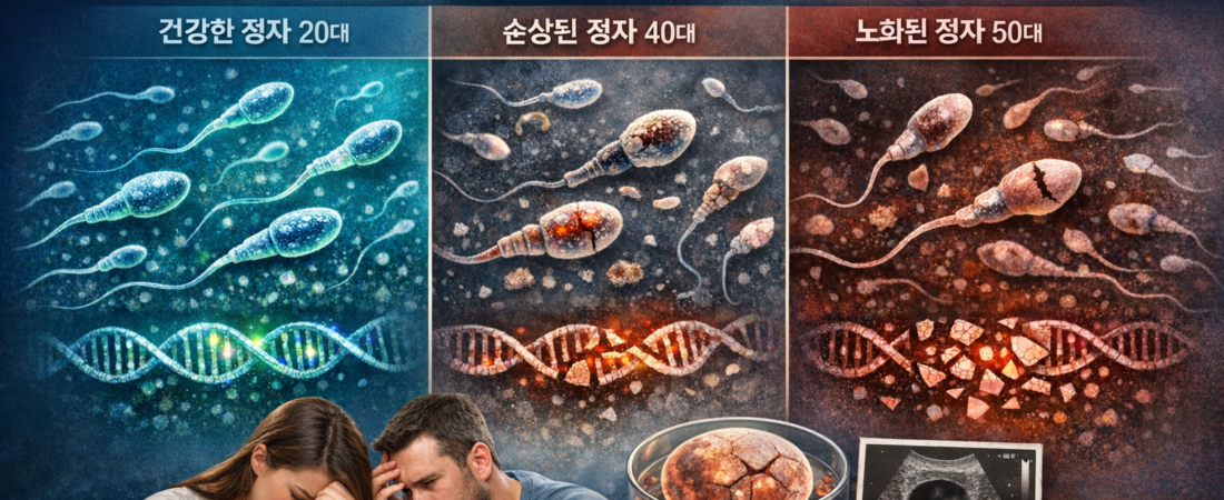
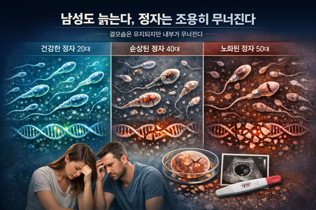
During a man’s 20s, sperm cells are structurally highly stable, exhibiting exceptionally low rates of DNA damage. This phase represents the peak of sperm motility and overall fertilization capacity. The reason natural conception occurs seamlessly during this decade is not merely because the individual is “young,” but because the genetic data tightly packed within each sperm head remains in its most pristine, uncompromised state. Sperm cells produced during this period display high uniformity and maintain highly stable cellular divisions once they form an embryo.
As men transition into their 30s, subtle shifts begin to materialize without warning. Sperm counts and motility parameters rarely present clear clinical abnormalities at this stage, but the rate of DNA damage begins a slow, progressive climb. Clinically, this decade is often marked by an increase in cases where couples face unexplained infertility despite receiving entirely “normal” routine semen analysis reports. Reproductive endocrinologists view this specific window as the initiation of invisible fractures—the exact phase where recurrent reproductive failures, unexplained by legacy testing, begin to manifest.
The 40s represent a definitive biological turning point. From this decade onward, declines in sperm motility and visible morphological abnormalities become clinically apparent. Most crucially, the DNA Fragmentation Index, or DFI, exhibits a statistically significant escalation. This molecular disruption does more than simply lower the mathematical probability of fertilization; even if fertilization is successfully achieved, the resulting embryo undergoes unstable development, directly driving up the clinical risk of early miscarriage. In active fertility clinics, a distinct diagnostic pattern of “successful fertilization followed by a failure to sustain gestation” becomes highly prominent during this decade.
From the 50s and beyond, the clinical conversation shifts entirely away from the quantity of sperm and focuses strictly on its intrinsic quality. Even if sperm concentrations remain numerically sufficient, the proportion of sperm cells carrying undamaged, healthy genetic data plummets. In other words, while the raw volume appears adequate on the surface, the pool of biologically viable sperm has radically diminished. At this advanced stage, a standard semen analysis loses its diagnostic utility; clinicians must comprehensively evaluate DNA damage, systemic oxidative stress, and the patient’s holistic metabolic profile to formulate an accurate assessment.
The absolute core of this biological reality distills into a single truth: while sperm cells are continuously manufactured, the cellular environment responsible for their production is steadily aging. This is not an isolated pathology of the testes. Systemic factors—including micro-vascular blood flow, endocrine balance, low-grade chronic inflammation, and even the gut microbiome—exert a direct, continuous influence on oocyte-fertilizing capacity. Ultimately, a sperm cell is less a direct consequence of chronological age and more the holistic sum of a man’s total systemic health.
Consequently, the diagnostic standards for male factor infertility are undergoing a profound clinical evolution. In the past, evaluations centered almost exclusively on visible, high-level metrics such as count, motility, and morphology. Today, invisible molecular markers—specifically DFI and oxidative stress levels—are treated with far greater clinical weight. The paradigm has shifted from a superficial question of “normal versus abnormal” to a deeper investigation into how safely and accurately a sperm cell can deliver unfragmented paternal genetic data to the egg.
This transition is not a minor medical update; it represents a fundamental shift in the reproductive paradigm. The cultural myth that a man can comfortably become a father at any point in his lifespan is no longer a scientifically viable hypothesis.
The accurate biological formulation is this: men age too. However, sperm degrades quietly. The damage occurs first in the microscopic spaces hidden from view, and the downstream consequences reveal themselves not through a simple lack of conception, but through the devastating cycles of IVF failure and miscarriage.
Editor’s Note: This commentary has been comprehensively reconstructed based on publicly available reproductive medicine literature, peer-reviewed international journals, and global health guidelines established by the World Health Organization (WHO). All medical diagnoses and clinical interpretations must exclusively be sought through direct consultation with a qualified fertility specialist.
Agarwal A, et al. “Sperm DNA fragmentation: a new guideline for clinicians.” World Journal of Men’s Health, 2020.
Esteves SC, et al. “Clinical relevance of sperm DNA fragmentation testing.” Andrology, 2021.
Evenson DP. “Sperm chromatin structure assay (SCSA) and DNA fragmentation.” Human Reproduction Update, 2016.
Sharma R, et al. “Effects of increased paternal age on sperm quality and fertility.” Reproductive Biology and Endocrinology, 2015.