
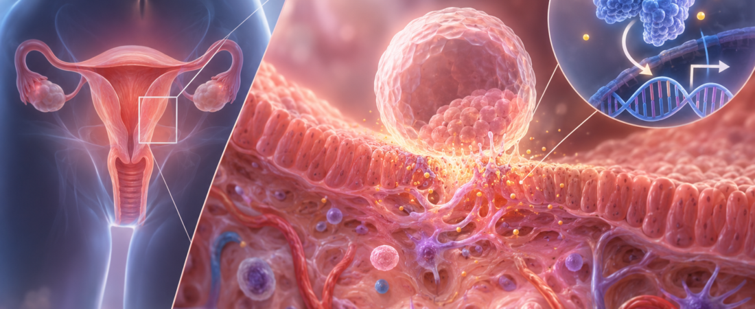
Why can implantation fail even after a good-quality embryo is transferred?
STAT3 and implantation research examines decidualization—the uterine lining’s transformation in preparation for pregnancy. The evidence includes animal experiments, human tissue studies, and cell experiments, not an established treatment.
“They told me it was a good embryo. Why did it fail to implant?”
It is one of the hardest questions to answer in in vitro fertilization (IVF). An embryo may develop well to the blastocyst stage and look good under the microscope. Even an embryo selected through preimplantation genetic testing for aneuploidy (PGT-A) because it has the expected chromosome number may fail to lead to pregnancy.
A good-quality embryo does not always implant.
Successful implantation requires more than a suitable embryo: the endometrium, or uterine lining, must also become ready to receive it. Among the many molecular signals involved in this process, researchers have been studying signal transducer and activator of transcription 3, or STAT3.
STAT3 is a protein that relays signals received from outside a cell and helps regulate the activity of particular genes. In the endometrium, it is involved in embryo implantation and decidualization.
How does the uterine lining change before an embryo arrives?
Implantation is not simply a matter of an embryo sticking to the surface of the uterine lining.
After ovulation, rising progesterone levels prompt endometrial stromal cells to change their shape and function. These changes create an environment that can receive an embryo and subsequently support pregnancy.
This process is called decidualization.
Decidualization changes not only the appearance of endometrial cells but also gene expression, the immune environment, and functions involved in blood-vessel formation. It must occur at the appropriate time to support embryo invasion into the lining and the beginnings of placental development.
An adequately thick lining therefore does not necessarily mean that every aspect of implantation readiness is in place. Beyond the thickness measured on ultrasound, complex molecular changes are occurring inside endometrial cells.
STAT3 is one of the signals involved.
What happened when uterine STAT3 was removed in mice?
One influential study of STAT3’s role was published in The FASEB Journal in 2013.
The researchers selectively removed STAT3 function in the mouse uterus and observed the consequences.
The absence of STAT3 did not produce major abnormalities in ovarian function or basic uterine development. However, implantation was disrupted: embryos failed to implant normally into the lining, and decidualization did not proceed properly.
In particular, signaling associated with the progesterone receptor was altered.
Progesterone is a key hormone that changes the uterine lining after ovulation to support pregnancy. The researchers explained that STAT3 helps regulate progesterone-receptor-related signaling and plays an important role in uterine implantation and decidualization.
This was an animal experiment, however. Its findings cannot simply be treated as an explanation for recurrent implantation failure in humans.
What differences, then, can be found in the uterine lining of women with recurrent implantation failure?
Lower STAT3 activity in women with recurrent implantation failure
The endometrium of women with recurrent implantation failure showed differences.
Could changes of this kind also be found in human endometrial tissue?
A study published in the 2021 volume of the Journal of Advanced Research examined endometrium collected during the implantation window from women with recurrent implantation failure.
The researchers focused on a chain of signals within cells as the uterine lining prepares for pregnancy. STAT3 was central to that pathway.
Compared with controls, women with recurrent implantation failure had lower levels of activated STAT3, known as phosphorylated STAT3 or p-STAT3. For STAT3, being present is not enough: it must be activated to perform its function. This activation signal was relatively weaker in the women with recurrent implantation failure.
Levels of interleukin-6 (IL-6), which participates in STAT3 activation, were also lower. Markers associated with decidualization—the lining’s transformation into a state suited to pregnancy—were reduced as well.
The researchers observed similar patterns in cell experiments. Suppressing the relevant signaling reduced STAT3 activity and impaired decidualization in endometrial cells.
The central finding is straightforward. In some women with recurrent implantation failure, STAT3 signaling was less active during the process of preparing the lining to receive an embryo, while decidualization also appeared impaired.
This suggests that repeated implantation failure after the transfer of good-quality embryos should not be investigated solely from the embryo’s perspective. Functional changes in the lining that receives the embryo also deserve attention.
Cell experiments also linked the pathway to decidualization
The researchers also carried out experiments using human endometrial cells.
When they suppressed signals involved in endometrial STAT3 activation, STAT3 activity fell and decidualization was impaired. Conversely, restoring the relevant signaling recovered some cellular functions.
They interpreted these findings as evidence that disruption of signaling leading to STAT3 could affect the process by which the lining prepares to receive an embryo.
STAT3 alone cannot explain recurrent implantation failure. Implantation is influenced by many factors, including the embryo’s chromosomes and developmental status, uterine structure, the condition of the endometrium, and hormones.
It is therefore more accurate to regard STAT3 as one of several signals being investigated to understand implantation readiness, rather than to label it the cause of recurrent implantation failure.
A thick lining may still differ in function
The endometrial measure most readily available to patients during fertility treatment is thickness.
Before embryo transfer, ultrasound measurements may show a lining of 7 mm, 8 mm, or 10 mm. This makes it easy to assume that adequate thickness means adequate preparation for implantation.
However, endometrial thickness is only one measure of the lining’s condition.
After ovulation, progesterone changes gene expression in the endometrium, stromal cells undergo decidualization, and the immune and vascular environments change. Many of these processes cannot be directly assessed with a routine ultrasound scan.
Two linings of similar thickness therefore need not provide identical implantation environments at the molecular level.
This is one reason STAT3 research is interesting: it offers a clue to the functional preparation for implantation that ultrasound cannot show.
Would increasing STAT3 improve implantation rates?
Could raising STAT3 activity make implantation more likely?
If a patient has weak STAT3 signaling, would strengthening that signal improve the implantation rate?
We do not yet know.
A recent cell study reported that granulocyte colony-stimulating factor (G-CSF), which has also been used in fertility treatment, may activate STAT3 and promote decidualization in endometrial cells. Blocking STAT3 reduced these effects.
This provides further experimental evidence that STAT3 may have an important role in preparing the lining for implantation.
It does not establish a treatment ready for routine use. Testing whether STAT3 is functioning properly is not currently a standard way to diagnose the cause of recurrent implantation failure. Nor has it been adequately demonstrated that treatment to increase STAT3 signaling improves pregnancy or live birth rates.
Showing that STAT3 participates in implantation-related processes in cultured cells is different from improving a patient’s chance of pregnancy by altering that pathway.
For now, STAT3 is best understood as an important research target for explaining implantation, rather than as a new implantation treatment.
Beyond a good embryo: a uterus ready to receive it
IVF technology has long advanced through efforts to identify good embryos. Embryo culture has improved, culture to the day-5 blastocyst stage has become commonplace, and PGT-A can be used to examine embryo chromosome numbers.
Yet not every embryo with the expected chromosome number implants.
Research is therefore looking beyond embryo quality to the biological changes that occur when an embryo and the uterine lining meet.
STAT3 and decidualization research is part of this wider effort.
STAT3 cannot explain recurrent implantation failure on its own, and manipulating it does not guarantee implantation. These studies do, however, show why pregnancy depends not only on the embryo’s characteristics but also on the endometrium undergoing appropriate decidualization at the right time and becoming ready to receive it.
Implantation does not end with selecting a good embryo and placing it in the uterus.
When the embryo arrives, the lining must also be ready to support pregnancy. STAT3 is being investigated as one important signal within this complex process.
For clinical context, read our explanation of how recurrent implantation failure is assessed and why unproven add-ons require caution.
Research sources
This article draws on the study of progesterone-induced blocking factor 1 (PIBF1), IL-6, p-STAT3, and endometrial decidualization in women with recurrent implantation failure, published in the 2021 volume of the Journal of Advanced Research (online September 9, 2020): DOI: 10.1016/j.jare.2020.09.002; the 2013 FASEB Journal mouse study of STAT3 and uterine implantation: DOI: 10.1096/fj.12-225664; and related subsequent research.
The G-CSF experiment discussed above used cultured human endometrial stromal cells, not a clinical pregnancy trial: “Granulocyte Colony Stimulating Factor Enhances Decidualization Process of Endometrial Stromal Cells Through STAT3/HOXA10 Axis,” Advanced Biology, 2025, DOI: 10.1002/adbi.202400279.
This article explains infertility and reproductive-medicine research. It does not replace individual diagnosis or treatment.
The image used in this article was created using generative artificial intelligence (ChatGPT, OpenAI). Any people depicted are fictional, not real individuals.
Copyright © 2026 Fertility Information TokTok Fertility News | THE FERTILITY NOTES. All rights reserved. Unauthorized republication, reproduction, redistribution, scraping, AI training, and data use of any content, including articles, photographs, and images, are strictly prohibited. Violations may result in legal liability.