

A microelectrode chip measures blastocyst oxygen use to quantify metabolic activity.
Mouse blastocyst hatching rates differed at 75% versus 11%; testing in 42 human blastocysts suggested potential, but did not assess pregnancy outcomes.
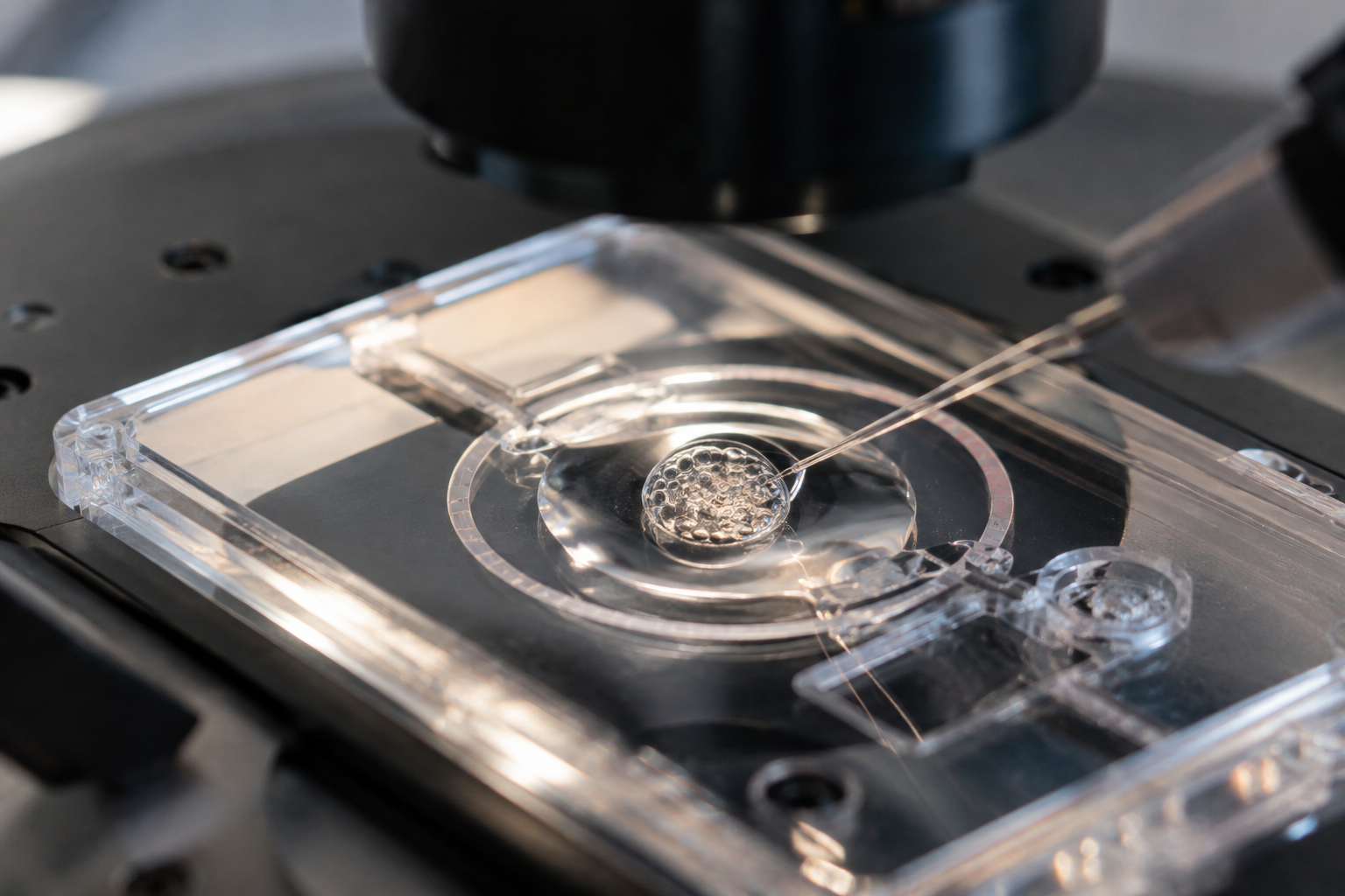
Choosing an embryo with a good chance of implantation is an important part of IVF. Traditionally, developmental potential has been assessed largely by observing embryo appearance and cellular structure under a microscope.
A new technique that measures how much oxygen an embryo consumes could add a different kind of information to embryo assessment.
In research published October 15, 2026 in Sensors and Actuators B: Chemical, investigators developed a microelectrode chip to measure blastocyst oxygen consumption precisely and examine its relationship with development and hatching.
In mouse embryos, a size-adjusted oxygen-consumption measure showed promise for distinguishing hatching potential beyond conventional morphological grading.
What embryo appearance cannot reveal
Morphological assessment remains widely used in IVF. Grading considers blastocyst expansion and development of the inner cell mass and trophectoderm.
Embryos with similar appearances can nevertheless differ in developmental potential. Morphology alone cannot fully describe internal metabolic activity or energy production.
The researchers focused on oxygen consumption. Developing embryos need energy, and mitochondria use oxygen to generate it. Oxygen use can therefore serve as an indirect measure of mitochondrial activity and metabolic state.
Measuring it is difficult because embryos are tiny and sensitive to environmental change. A change in embryo position or culture-medium conditions can influence the result.
The team developed a microelectrode chip that holds a blastocyst steadily in a defined position while measuring changes in surrounding oxygen concentration.
How the oxygen-sensing microchip works
The device integrates a concentric stepped microstructure, oxygen-sensing electrodes and a salt-bridge channel on one chip. With the blastocyst positioned in place, nearby electrodes detect changes in dissolved oxygen, from which oxygen consumption is calculated.
Measurements were performed at 37°C to reflect embryo-culture conditions. An oil layer limited evaporation. Intermittent measurement consisted of 40 seconds of oxygen sensing followed by a 260-second recovery period.
The reference electrode also showed stability, with a potential change of approximately 1 millivolt over 30 minutes.
The design allowed an embryo to be held steadily and retrieved when needed, suggesting possible future use in embryology laboratories.
However, the study demonstrated measurement feasibility under experimental conditions. It did not fully establish the effects of repeated measurement on human embryo development.
Mouse hatching rates: 75% versus 11%
The researchers measured oxygen consumption in 17 mouse blastocysts and examined its relationship with development.
The key measure was not simply total oxygen use, but consumption adjusted for embryo size. Because larger embryos may consume more oxygen, the team divided oxygen-consumption rate by embryo radius.
This adjusted measure distinguished good-grade blastocysts from other blastocysts more effectively than oxygen-consumption rate alone.
It also related to hatching. Blastocysts at or above the reported size-adjusted threshold of 0.43 fmol/(s·μm) had a hatching rate of 75%, compared with 11% below it.
Hatching is the process in which a blastocyst escapes the surrounding zona pellucida, a developmental step preceding implantation in the uterine lining.
The result supports further investigation of oxygen consumption as a marker of developmental potential. It came from a small experiment involving 17 mouse embryos and must not be interpreted as a human pregnancy-success rate.
Findings in 42 human blastocysts need clinical validation
The investigators also measured oxygen consumption in 42 human blastocysts. The size-adjusted measure showed potential for distinguishing morphologically good-grade from fair-grade blastocysts.
This suggests that the technique could quantify aspects of metabolic state in human embryos as well as mouse embryos.
However, the study did not transfer human embryos and compare clinical pregnancy or birth outcomes. It cannot establish that selecting human embryos with higher oxygen consumption improves pregnancy or the chance of a healthy birth.
Oxygen consumption is only one metabolic measure. It does not establish chromosomal normality or explain every aspect of implantation potential. Larger human-embryo studies need to confirm reproducibility and link measurements with actual transfer outcomes.
From appearance toward physiological function
The study’s contribution is to extend embryo assessment from visual observation toward measurement of physiological function.
Morphology, time-lapse imaging and AI-based assessment are used or being investigated in IVF. These primarily analyze appearance and developmental patterns, whereas oxygen-consumption measurement seeks to quantify metabolic activity.
Combining such measures with existing methods could eventually provide a more multidimensional assessment. Metabolic differences between similarly graded embryos might offer useful information for research and evaluation.
At present, the technology has not been shown to replace embryo grading or improve pregnancy-success rates.
Its significance lies in looking beyond how an embryo appears to what it is doing metabolically. Clinical evidence incorporating human embryo transfer and birth outcomes will be needed to establish its contribution to embryo selection.
Source: This article is based on “A dissolved oxygen electrode chip with concentric stepped microstructure and integrated salt-bridge channel for blastocyst positioning and oxygen consumption rate measurement,” published October 15, 2026 in Sensors and Actuators B: Chemical. Authors include Ping-Feng Yang, En-Hui Cheng, Pin-Chi Tang, Hitoshi Shiku, Maw-Sheng Lee and Ching-Chou Wu. The study examined 17 mouse and 42 human blastocysts. DOI: 10.1016/j.snb.2026.140135.
Image disclosure: The image was created using generative AI (ChatGPT, OpenAI); any people depicted are fictional, not real individuals.
Related reading on assessing evidence before adopting additional IVF techniques: what a review of IVF add-ons found about live birth.
Copyright © 2026 Fertility Information TokTok Fertility News | THE FERTILITY NOTES. All rights reserved. Unauthorized reproduction, copying, redistribution, scraping, AI training, and other data use of any articles, photographs, images, or other content are strictly prohibited. Violations may result in legal liability.