
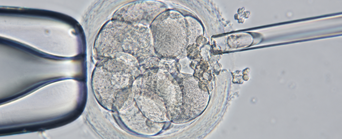
Embryo fragmentation in IVF can leave small pieces of cytoplasm between developing cells. The DEBRIS randomized trial is testing whether removing those fragments on day 4 improves blastocyst development and later pregnancy outcomes. The planned enrollment is 320 participants. This is a study of a possible treatment, not evidence that defragmentation already improves IVF success.
Embryos with at least 10% fragmentation are being studied. The technique uses a laser to open the zona pellucida and a fine needle to remove fragments. Blastocyst development and pregnancy outcomes will be compared; a clinical benefit has not yet been established.
During embryo culture in in vitro fertilization, small pieces of material can appear between the cells. This is called embryo fragmentation. If these pieces are deliberately removed, could an embryo be more likely to grow into a blastocyst?
A randomized controlled trial is investigating that question by removing cytoplasmic fragments on the fourth day after fertilization, then assessing embryo development and pregnancy outcomes.
What are the fragments seen between embryo cells?
A fertilized egg divides from one cell into two, four, eight and progressively more cells. That process does not always proceed neatly. Small pieces of cytoplasm without a nucleus can remain between the dividing cells. These are the cytoplasmic fragments that look like tiny pieces under the microscope.
Embryologists have long assessed the extent of fragmentation alongside cell number, cell size and the pace of division when evaluating embryo morphology.
That raises an apparently simple question: if the fragments are a problem, why not remove them?
This is the idea behind embryo defragmentation. Attempts have previously been made in early embryos on days 2–3 after fertilization. However, inconsistent findings have prevented the technique from becoming a standard IVF treatment.
The current trial focuses on a less studied time point: day 4, when the embryo’s cells are compacting as it moves toward the morula stage.
How day-4 embryo defragmentation is performed
The protocol describes embryos assessed about four days after fertilization—specifically 96 ± 2 hours after ICSI—with at least 10% cytoplasmic fragmentation.
The procedure is technically precise. A diode laser with a wavelength of 1.48 micrometers creates a small opening in the zona pellucida, the outer coat surrounding the embryo. A 35-micrometer biopsy needle is then used to aspirate fragments from between the cells.
Softer fragments are aspirated directly. Firmer fragments are anchored with the needle and mechanically extracted. Manipulation takes place on a heated stage at 37°C, with a maximum manipulation time of 10 minutes per patient. The embryos then return to the incubator for culture to the blastocyst stage on days 5–6.
Will more embryos reach the blastocyst stage?
The trial compares embryos undergoing fragment removal with those continuing standard culture without additional manipulation. For the randomized comparison, patients with day-4 embryos showing at least 10% fragmentation are assigned to the intervention or standard-culture group. Embryos with less than 10% fragmentation provide a biological reference group.
The investigators want to determine whether treated embryos are more likely to reach the blastocyst stage on days 5–6, and whether any laboratory improvement is accompanied by better pregnancy outcomes.
A recruiting trial is not a proven treatment
The idea is interesting, but an essential distinction must be maintained. This is not a completed study demonstrating that removing embryo fragments increases pregnancy rates. It is a clinical trial designed to find out whether such an effect exists.
Additional manipulation of an embryo cannot be assumed to be risk-free. Because fragment removal involves physical intervention, a randomized comparison is needed to establish whether any benefit outweighs the risks of that manipulation.
Are the fragments merely a visible sign that an embryo is developing less well? Or do the fragments themselves interfere with further development, so that removing them could help restore developmental potential?
Results from this planned 320-participant trial may provide useful evidence on whether directly removing the fragments that embryologists have generally observed under the microscope offers a real benefit.
Study source, design and current status
This article is based on the public protocol for the prospective randomized trial registered as NCT07500337, DEBRIS, sponsored by Momo Fertilife. The registered study investigates the effect of day-4 embryo defragmentation on blastocyst development and clinical outcomes in ART cycles.
The registry was checked on September 29, 2026: the study was recruiting, its estimated enrollment was 320, and no results were posted. The protocol uses patient-level randomization and blinded outcome assessment. Its planned outcomes must not be presented as findings, and pregnancy or live-birth benefits remain unproven.
Related reading
- Embryologists in IVF: Skill, Checks and Laboratory Safety
- Direct Embryo Warming: Promising Mouse Data, Human Limits
Images were created with generative AI (ChatGPT, OpenAI) for educational illustration and are not clinical photographs of the study participants.
Copyright © 2026 Fertility Information TokTok | THE FERTILITY NOTES. All rights reserved. Unauthorized republication, reproduction, redistribution, scraping, AI training, or other data use of any content, including articles, photographs and images, is strictly prohibited. Violations may result in legal liability.