
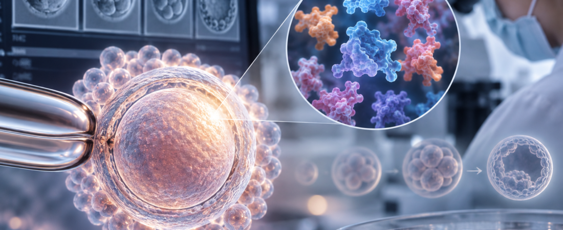
Egg quality in IVF cannot be read from appearance alone. Researchers studying the oocyte proteome found a protein signature associated with developmental competence in mouse eggs, then confirmed key proteins in 49 human oocytes. The work points toward more objective assessment, but it has not produced a clinical test that selects eggs likely to lead to pregnancy.
The October issue of Human Reproduction reports differences in protein synthesis, autophagy and intracellular transport across eggs with different developmental potential. Approximately 1,700 proteins were identified in oocytes, and key candidates were also examined in donated human eggs.
Patients undergoing IVF often hear the phrase “egg quality.” Eggs retrieved on the same day may have very different outcomes: some fertilize and reach the blastocyst stage, while others do not fertilize or stop developing. What, then, makes a good egg different?
Assessment has traditionally depended on morphology: examining shape and maturity under a microscope. Yet mature eggs that look alike do not necessarily have the same capacity to support development after fertilization.
A recent study sought that difference in an egg's internal “protein fingerprint.” Identifying protein combinations associated with fertilization and embryo growth could eventually help researchers understand egg quality more objectively.
Researchers at UNSW Sydney in Australia published the comparison in the October 2026 issue of Human Reproduction, pages 1741–1761, following online publication on August 20.
Eggs with different developmental potential had different proteomes
The study focused on oocyte developmental competence: the capacity to fertilize normally and support subsequent embryo development.
The investigators compared mouse oocytes under three maturation conditions: eggs matured in the body, eggs matured using conventional in vitro maturation (IVM), and eggs matured with CAPA-IVM, which regulates maturation in stages.
Mass spectrometry was used to examine the proteins and their abundance in the eggs and their surrounding cumulus cells.
The researchers identified 1,736 proteins in oocytes and 3,212 in cumulus cells. After stricter analytical filtering, approximately 1,600 oocyte proteins and 3,100 cumulus-cell proteins remained in the comparisons.
Overall protein composition differed clearly according to how the eggs matured.
In oocytes, differences involved translation, the process of making proteins; autophagy, which clears unwanted or damaged cellular material; and endocytosis, which helps cells take up and transport material. Cumulus-cell changes included pathways involved in reactive-oxygen-species detoxification and serine biosynthesis.
In everyday terms, a good egg may be more than an egg that looks attractive under a microscope. Its internal systems for producing, clearing and transporting proteins may be in a different state as it prepares for early embryo development.
Clathrin, RPL24 and EIF2A emerged as candidate proteins
The researchers then used additional methods to test whether selected differences from the large-scale protein analysis could be confirmed.
Changes involving clathrin, which participates in intracellular transport; ribosomal protein L24 (RPL24); and eukaryotic initiation factor 2A (EIF2A), involved in protein synthesis, were consistent with the proteomic findings.
These proteins are not simply passive contents of an egg. They are linked to basic cellular activities required to produce proteins and move materials to the right places as an egg transitions toward an early embryo after fertilization.
The findings suggest that developmental competence may reflect several molecular systems working together, rather than a single visible feature.
The team also examined 49 human oocytes
The work did not end with mouse experiments. The researchers used 49 human oocytes donated for research by 36 patients receiving assisted reproductive technology (ART) treatment to investigate whether key proteins identified in mouse eggs were also present in human eggs.
Some oocytes underwent rescue IVM, allowing protein expression to be examined in immature and mature MII oocytes.
Clathrin, RPL24 and EIF2A were identified in human GV-stage immature eggs and mature MII eggs. This suggests that some protein features observed in mice may be conserved in humans.
Can a protein test now select a “good egg”?
A clear distinction is needed: the study did not establish that a protein test can identify eggs that will produce successful pregnancies. The central proteomic analysis was performed in mouse oocytes. The human component confirmed the presence of candidate proteins.
The researchers also noted that human MII oocytes can differ in quality even within the same patient and retrieval cycle, making the mechanisms underlying human developmental competence difficult to unravel.
Directly analyzing an egg's proteins may also create practical problems if that egg is then needed for fertilization and transfer. Clinical application would therefore require ways to assess quality indirectly without damaging the egg.
That makes the surrounding cumulus cells important. Their protein changes involved reactive-oxygen-species detoxification, serine synthesis, and steroid and cholesterol biosynthesis.
If surrounding cells can provide information about the egg without manipulating it directly, they might eventually support noninvasive assessment. That possibility still requires additional human studies and clinical validation.
From “how many eggs?” to “what kind of eggs?”
Egg number is important in IVF, but it does not explain everything. People with the same number of retrieved eggs can have different fertilization rates, blastocyst formation and pregnancy outcomes. Differences in developmental competence are one reason.
This study's contribution is not an immediately available egg-selection test. It extends assessment beyond microscopic appearance to the molecular processes that may need to function properly for an egg to have strong developmental potential.
The authors suggest that defining these molecular features could contribute to future diagnostics that quantify developmental competence and help improve assisted reproduction.
Study source
This article is based on “A proteomic signature of oocyte quality from models of varying oocyte developmental competence,” by Emily R. Frost, Dulama Richani, Robert B. Gilchrist and colleagues at the Fertility & Research Centre, UNSW Sydney, Australia. Published online August 20, 2026, and in Human Reproduction 41(10):1741–1761, October 2026. PMID: 42625370. DOI: 10.1093/humrep/deag134. Proteomics data: ProteomeXchange PXD073269.
Related reading
Rescue IVM and the developmental potential of immature eggs
The illustration was generated using ChatGPT (OpenAI). It is not a clinical image or a photograph of actual patients.
Copyright © 2026 THE FERTILITY NOTES. All rights reserved. Unauthorized republication, reproduction, redistribution, scraping, AI training and data use of any content, including articles, photographs and images, are strictly prohibited. Violations may result in legal liability.