
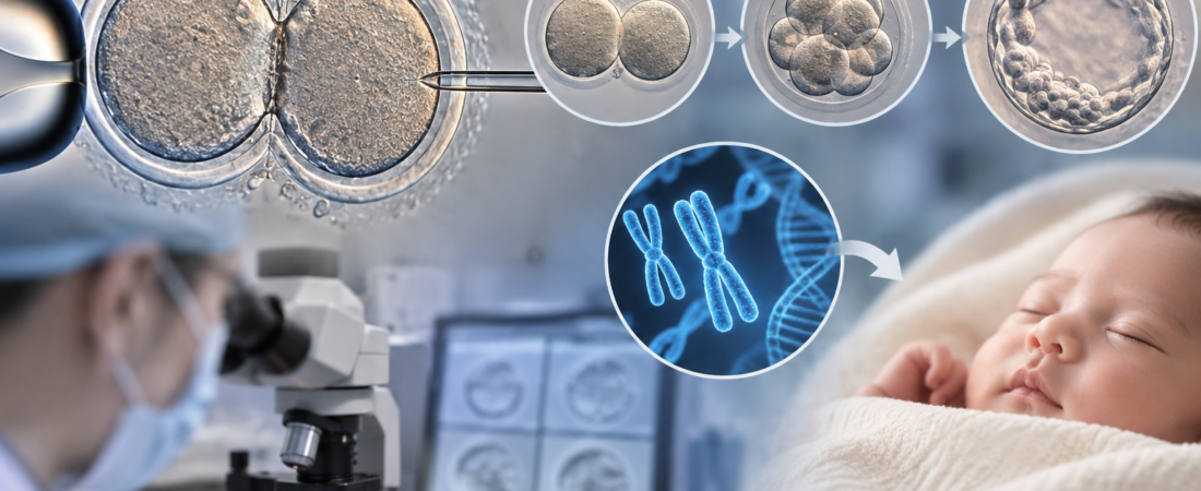
Conjoined oocytes in IVF can look startling: two eggs appear attached to each other. A study of 42 pairs found lower maturation and fertilization rates, but embryos that cleared fertilization could develop into blastocysts, including some with euploid PGT-A results. The study did not establish pregnancy or live birth safety.
In 39 treatment cycles, maturation was 36.9% and fertilization was 48.38%. After fertilization, development did not differ significantly from ordinary oocytes; five of eight tested blastocysts were euploid. The authors see a basis for considering their potential when no other embryos are available, while calling for further pregnancy and birth research.
During an IVF egg retrieval, an embryologist may occasionally see two connected eggs rather than a single oocyte. These rare conjoined oocytes look different from ordinary eggs, and evidence has been limited on whether they can be fertilized and develop into normal embryos.
An unusually substantial series of these rare eggs produced an interesting finding. Conjoined oocytes were less likely to mature and fertilize, but once they passed the first hurdle of fertilization, their ability to form embryos and blastocysts was not clearly inferior. Five of eight blastocysts undergoing preimplantation genetic testing for aneuploidy (PGT-A) had a normal chromosome number.
Two eggs from one follicle, attached at the zona pellucida
Researchers at the Affiliated Yantai Yuhuangding Hospital of Qingdao University in China analyzed 39 IVF/ICSI cycles in 38 patients with conjoined oocytes between January 2012 and September 2025. They identified 42 pairs, or 84 individual oocytes. The findings were published in Reproductive Biology and Endocrinology on September 29, 2026.
The investigators defined conjoined oocytes as two eggs originating from the same follicle and attached at the zona pellucida, the outer coat surrounding an egg. This is a very rare departure from the usual development of one egg per follicle.
Being attached does not mean that both eggs mature normally together.
Only 31 of 84 oocytes matured
Of the 84 conjoined oocytes, 31 reached the mature metaphase II (MII) stage: a maturation rate of 36.90%, significantly lower than for ordinary oocytes.
Their developmental timing was often out of step. In 71.43% of pairs, one egg had reached MII while the other remained at the immature germinal-vesicle (GV) stage. The researchers identified this asynchronous maturation as a potentially important contributor to reduced fertilization potential.
Size also differed. The larger egg in a pair had approximately twice the area of the smaller one, but both were smaller than ordinary oocytes. The zona pellucida around the smaller egg averaged 12.05 micrometers thick, thinner than around the larger partner and ordinary eggs.
Fertilization was harder—but development told a different story
Fifteen of the 31 mature conjoined oocytes fertilized normally, a fertilization rate of 48.38%, significantly below the comparison group.
That initially looks discouraging. Continued culture, however, revealed a different pattern.
After fertilization, the proportions developing into high-quality day-3 embryos, usable blastocysts and high-quality blastocysts did not differ significantly from ordinary oocytes. The results suggest that the major obstacle may be reaching normal fertilization; early development after that point may be more comparable. A nonsignificant difference does not establish equivalence.
Five of eight PGT-A blastocysts were euploid
The chromosome findings were particularly striking. Of eight blastocysts derived from conjoined oocytes that underwent PGT-A, five were classified as euploid, meaning a normal chromosome number. That is 62.5% by simple calculation.
The percentage must not be treated as a general euploidy rate for conjoined oocytes. Only eight blastocysts were tested. The findings neither show that they outperform ordinary eggs nor establish a reliable 62.5% rate for other patients.
The narrower implication is that an unusual, attached appearance alone is insufficient reason to assume that every resulting embryo has abnormal chromosomes.
Does this mean conjoined oocytes need not be discarded?
The authors remain cautious. This study did not follow transfers of the resulting embryos through pregnancy and birth. It cannot show that conjoined-oocyte embryos have the same pregnancy or live birth rates as embryos from ordinary eggs. The authors describe the evidence as hypothesis-generating and call for clinical outcome studies.
Earlier medical case reports have described healthy births after transferring blastocysts derived from conjoined oocytes. A 2016 report described a healthy girl after transfer of a chromosomally normal blastocyst, and subsequent reports described births following ICSI and frozen embryo transfer.
Those reports do not make conjoined eggs equivalent to ordinary eggs. Together with the new study, they add to the argument that unusual morphology alone may not justify assuming there is no developmental potential.
This could matter particularly when very few eggs are retrieved or no other usable normal embryos are available. The authors suggest that transfer of a resulting blastocyst might be considered when no other viable embryos exist. More chromosome analyses and longer follow-up of pregnancy, miscarriage and birth outcomes are needed before the findings can be applied more broadly.
Study source
This article is based on “Fertilization and developmental characteristics of conjoined oocytes in IVF/ICSI: a detailed analysis,” published September 29, 2026, in Reproductive Biology and Endocrinology. Authors: Guangzhong Jiao, Jianhua Wang, Lili Chen, Yinghua Shan, Hua Xing, Shuyuan Yin, Hongchu Bao, Xunlan Yin and Xiaoyan Liu, Department of Reproductive Medicine, Affiliated Yantai Yuhuangding Hospital of Qingdao University, China. DOI: 10.1186/s12958-026-01613-1.
Related reading
Rescue IVM for immature GV eggs
The illustration was generated using ChatGPT (OpenAI). It is not a clinical image or a photograph of actual patients.
Copyright © 2026 THE FERTILITY NOTES. All rights reserved. Unauthorized republication, reproduction, redistribution, scraping, AI training and data use of any content, including articles, photographs and images, are strictly prohibited. Violations may result in legal liability.