
Recurrent miscarriage and recurrent implantation failure may leave clues in the placenta even after a successful birth. A Mayo Clinic study of 522 women found differences in placental immune cells and tissue findings associated with those histories—but it did not establish a cause of pregnancy failure or a benefit from immune treatment.
Research into failed implantation and repeated miscarriage has often focused on embryo chromosomes, the endometrium and hormones. This study examined a different setting: placentas from pregnancies that continued to birth.
The investigators compared 325 women who conceived through IVF with 197 who conceived spontaneously. Placental inflammatory changes, blood-flow-related abnormalities and increases in selected immune cells were observed in association with previous recurrent pregnancy loss or implantation failure.
The distinction matters: these were placentas after successful pregnancies, not uterine samples collected at the time of failed implantation.
The placenta is also an immune interface
The placenta delivers oxygen and nutrients to the fetus and removes waste. It is also an important boundary between maternal and fetal immune environments.
A fetus inherits approximately half its genetic information from its father. Maintaining pregnancy requires finely balanced immune regulation that tolerates fetal tissues while retaining protection against infection.
Macrophages help remove damaged tissue and unwanted cells and regulate inflammation and repair. They also contribute to placental development and its immune environment.
The study examined macrophage markers CD68 and CD163. CD68 is widely used to identify macrophages, while CD163 helps characterize particular macrophage populations.
CD163-positive macrophages stood out after implantation failure
CD163-positive macrophages were particularly prominent in placentas from women with a history of recurrent implantation failure. Increases in CD68- and CD163-positive cells were also identified in IVF pregnancies with recurrent pregnancy loss or implantation-failure histories.
This suggests differences in the placental immune environment associated with previous reproductive failures.
More CD163-positive cells does not mean the immune system attacked the fetus. Although this marker is often associated with macrophage characteristics involved in inflammatory regulation or repair, function depends on the surrounding tissue environment.
The finding is an association between reproductive history and cell distribution, not evidence that an increase in these cells caused earlier pregnancy failures.
Vascular lesions and chronic inflammation
IVF placentas showed patterns of more frequent lesions suggesting impaired fetal-side vascular flow, chronic villitis and intervillous thrombi. These changes were particularly notable in women with recurrent pregnancy loss.
Chronic villitis involves inflammatory cells within the placental villi, structures important for exchange between mother and fetus. Inflammation and vascular abnormalities there can provide clues about placental function.
The IVF group with recurrent pregnancy loss showed a trend toward increased preeclampsia. Postpartum hemorrhage was statistically more frequent in the IVF group without recurrent pregnancy loss or implantation-failure histories.
Interleukin-6 (IL-6), an inflammation-related molecule, did not differ significantly between groups. Not every inflammatory measure moved in the same direction.
More immune cells does not establish a need for immune therapy
Although the overall comparison involved 522 women, placental histology was available for 283, and immunohistochemistry was performed on 133 specimens. The recurrent pregnancy loss and implantation-failure groups included 52 and 30 women, respectively, limiting subgroup analyses.
Because the placentas came from pregnancies that reached birth, the study cannot establish whether the cell changes existed before earlier failures or developed during the subsequent pregnancy.
Increased CD163- or CD68-positive cells therefore cannot establish the effectiveness of immunosuppressants, steroids or immunoglobulin. These placental markers are not established standard tests for diagnosing the cause of recurrent implantation failure or choosing treatment.
A research clue, not a confirmed explanation
The study broadens attention from embryos and the endometrium to the immune environment of the placenta that develops after implantation.
Finding differences after successful births suggests that a continuing pregnancy does not mean every placenta has identical immune-cell distributions or vascular characteristics.
The results may inform research into pregnancy monitoring and placental health after recurrent reproductive failure. Larger studies and investigations of cell function are needed before clinical applications can be established.
This work identifies a possible biological connection worth studying; it does not settle why a particular pregnancy failed.
Study source
SiWon Lee, SuYun Ling, Jade Rechtzigel, Chandra C. Paff Shenoy and Sylvie Girard, Mayo Clinic: “Pregnancy Outcomes, Placenta Histopathology and Immunohistochemistry in Patients with a History of Recurrent Pregnancy Loss or Implantation Failure Undergoing In Vitro Fertilization,” F&S Science, October 8, 2026. DOI: 10.1016/j.xfss.2026.09.004. PMID: 42849877.
Related reading
For context on assessing repeated transfer failures and additional tests, see our guide to recurrent implantation failure after euploid embryo transfer.
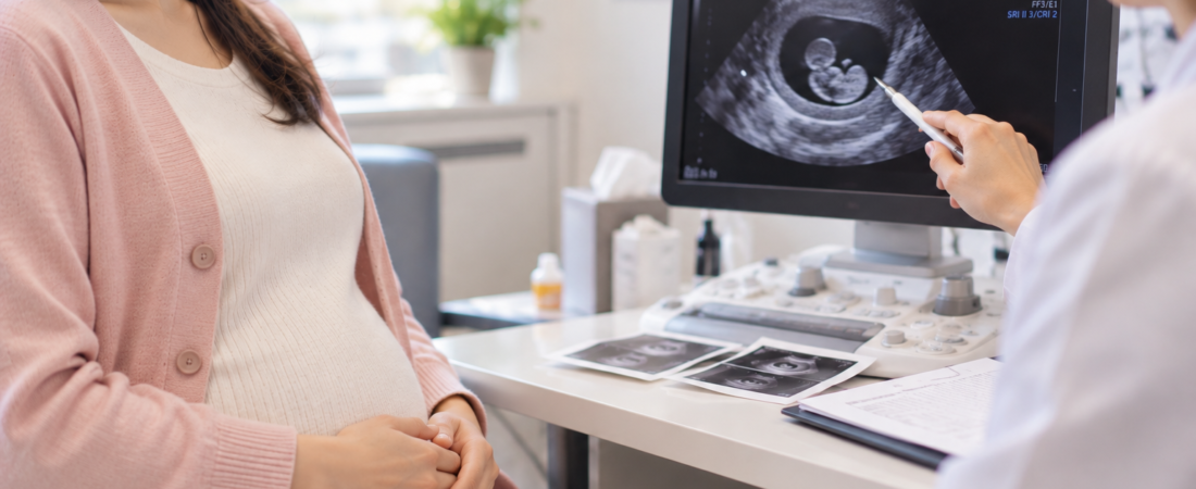
Illustration created with generative AI (ChatGPT, OpenAI); it does not depict an actual patient.
Copyright © 2026 The Fertility Notes. All rights reserved. Unauthorized reproduction, redistribution, scraping, AI training, or other data use of articles, photographs, images, and all other content is strictly prohibited and may result in legal liability.