
By Seungjoo Lee
Blastocyst collapse during IVF can look alarming: an expanded embryo suddenly contracts and may later expand again. A time-lapse study of 276 blastocysts found associations between collapse, embryo morphology and biochemical pregnancy. It also examined cytoplasmic strings, which showed a different pattern. Neither feature alone determines whether an embryo can produce a pregnancy or live birth.
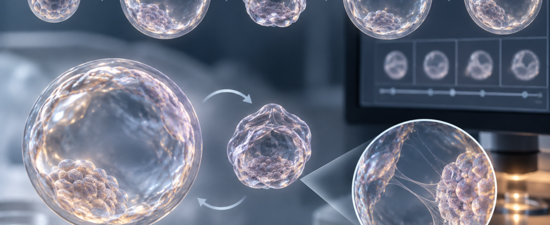
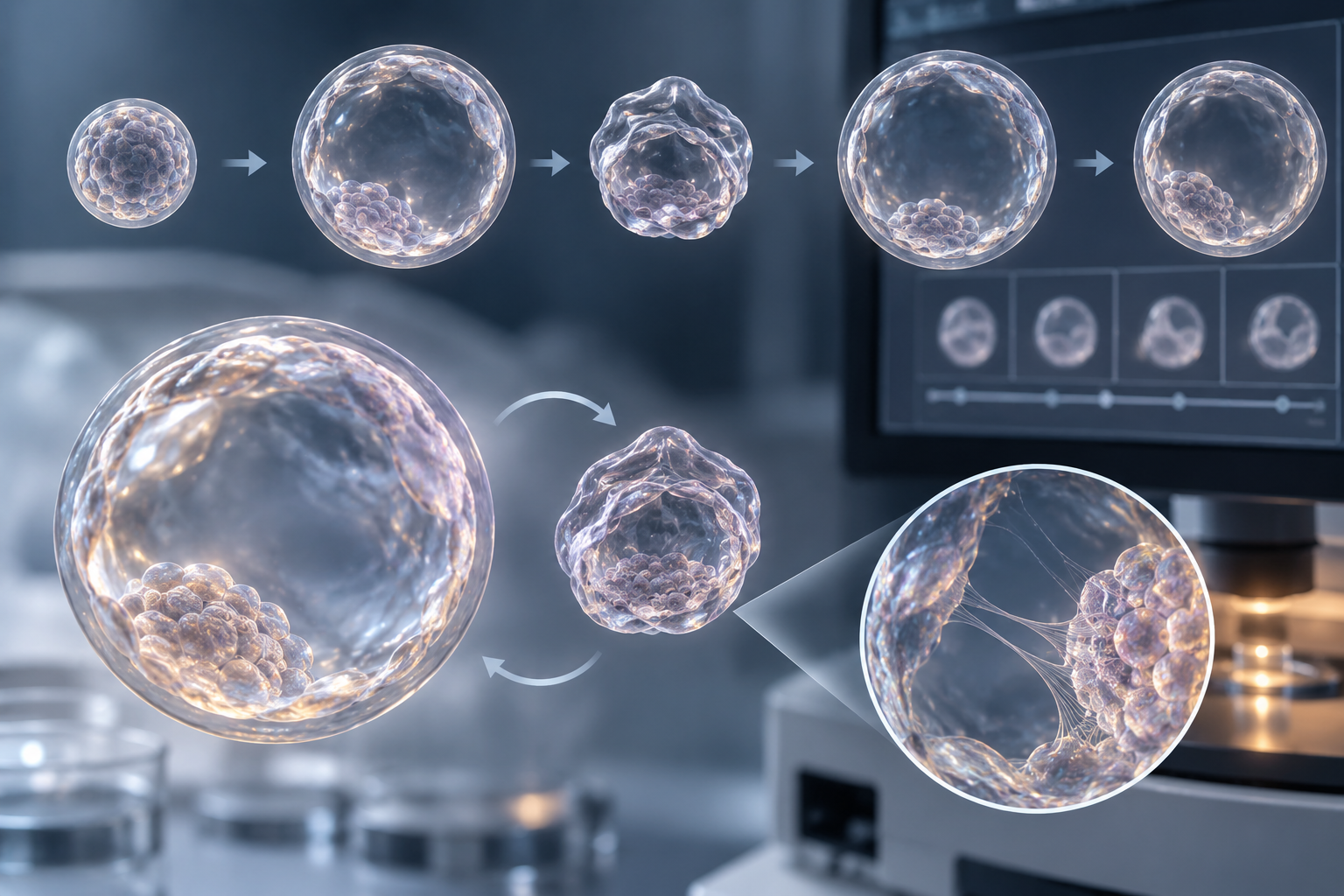
Watching an embryo in a time-lapse incubator can reveal surprising movements. A blastocyst that looked well expanded may suddenly shrink like a deflating balloon, then inflate again. This is known as blastocyst collapse.
At around five to six days after fertilization, fluid accumulates within the embryo and a cavity forms. The inner cell mass, which contributes to the fetus, becomes distinct from the trophectoderm, which contributes to the placenta. Occasionally, that expanding cavity abruptly becomes smaller and the whole embryo contracts.
Does this mean the embryo is in poor condition? Recent research suggests that movement during culture, alongside an embryo’s appearance, may offer another clue to developmental competence.
Watching 276 blastocysts with time-lapse imaging
Researchers at the Assisted Reproductive Technology Centre of Careggi University Hospital and the University of Florence, Italy, analyzed 276 blastocysts from 177 couples treated between 2022 and 2024.
They focused on two features: collapse during expansion and fine structures called cytoplasmic strings. These strings are thin filaments connecting the inner cell mass and trophectoderm.
Traditional morphological assessment grades an embryo’s appearance at particular moments. Time-lapse incubators take images at regular intervals without removing embryos, recording when they divide, become blastocysts, expand, contract and re-expand. This makes it possible to assess a sequence of movements rather than isolated snapshots.
Collapse occurred in approximately one in three blastocysts
Collapse was observed in 34% of the blastocysts. It was not a rare event, but its frequency differed by morphological grade: 14% of top-quality blastocysts, 29% of good-quality blastocysts and 43% of poor-quality blastocysts collapsed.
Thus, collapse was more common in embryos with poorer morphological assessments. The β-hCG-positive rate was 30% after transfer of blastocysts with collapse and 39% for those without collapse; the researchers reported a statistically significant difference.
β-hCG, or beta human chorionic gonadotropin, is the pregnancy hormone measured in blood after embryo transfer. It begins to be produced after implantation, and a positive result is an early marker used in assessing biochemical pregnancy.
The timing of collapse also mattered: it was inversely associated with β-hCG levels. This raises the possibility that an embryo’s changing shape supplies morphodynamic information about its developmental state.
Why can a well-expanded blastocyst suddenly shrink?
A blastocyst is not a motionless ball of cells. Trophectoderm cells regulate ion and water movement, drawing fluid into the blastocoel cavity and expanding the embryo.
Changes in internal pressure or connections between cells can allow fluid to escape, producing sudden contraction. When fluid accumulates again, the blastocyst re-expands. A single contraction therefore does not automatically mean that the embryo has died or stopped developing.
Collapse may nevertheless be associated with cellular structure, energy status or the stability of cell-to-cell connections. Earlier studies have reported less favorable implantation or ongoing-pregnancy outcomes among collapsing blastocysts. A 2020 time-lapse study defining collapse as a volume reduction of at least 50% reported higher implantation and ongoing-pregnancy rates among non-collapsing blastocysts.
Other research has associated collapse with higher amounts of cell-free mitochondrial DNA in culture medium, with more collapse events tending to accompany more cf-mtDNA. These findings do not, however, establish the complete biological mechanism behind collapse.
Cytoplasmic strings showed the opposite association
Cytoplasmic strings were observed in 61% of blastocysts: 71% of top-quality embryos, 57% of good-quality embryos and 32% of poor-quality embryos.
The β-hCG-positive rate was 76% for blastocysts with strings versus 51% for those without. The investigators proposed that collapse and strings might act as complementary non-invasive biomarkers, providing information in opposite directions about embryo competence.
The broader idea is to look beyond how an embryo appears at one moment. Its expansion and contraction during culture, and the structures formed by its cells, could add information about its development.
A collapsed blastocyst is not automatically a non-viable embryo
The findings should not be interpreted as “the embryo shrank once, so pregnancy is impossible.” This study of 276 blastocysts from 177 couples did not prove that collapse directly causes pregnancy failure. Positive β-hCG results occurred after transfer of collapsing blastocysts, while absence of collapse did not guarantee pregnancy.
Maternal age, embryo chromosomes, developmental speed, morphology, the inner cell mass and trophectoderm, endometrial conditions and transfer circumstances all contribute to outcomes. The β-hCG result highlighted here is an early pregnancy measure; it must not be treated as equivalent to live birth.
Morphology and developmental stage remain central to embryo selection, alongside other information such as PGT-A when indicated. Collapse or strings alone are not an established basis for deciding which embryos to transfer or discard.
Embryos continually change shape as they grow in the laboratory. Time-lapse technology captures movements that intermittent microscopic observation could miss. If these subtle movements can be integrated with morphology and chromosomal information, they may eventually help assess developmental potential without damaging an embryo. A brief contraction may be a biological signal worth studying, rather than a definitive verdict on the embryo.
Study source and limitations
Rossella Fucci, Asia Iserani and colleagues at Careggi University Hospital and the University of Florence, Blastocyst collapse and cytoplasmic strings as complementary morphodynamic biomarkers of embryo competence and biochemical pregnancy, Reproductive Biology, online September 22, 2026. Time-lapse analysis of 276 blastocysts from 177 couples receiving homologous assisted reproduction in 2022–2024; outcomes evaluated included embryo morphology, β-hCG positivity, miscarriage and live birth. The observational associations highlighted in this article do not establish causation or individual embryo prognosis. DOI: 10.1016/j.repbio.2026.101289; PMID 42772227.
This article introduces recent research and cannot determine an individual embryo’s suitability for transfer. The clinical and embryology teams must assess morphology, development and the patient’s circumstances together.
Related reading
Images were created using generative AI (ChatGPT, OpenAI) for illustration and do not depict actual patients.
Copyright © 2026 Fertility Information TokTok | THE FERTILITY NOTES. All rights reserved. Unauthorized republication, reproduction, redistribution, scraping, AI training and data use of articles, photographs, images and all other content are strictly prohibited. Violations may result in legal liability.