
“Why Does Implantation Fail Even with a 10mm Endometrium?”… The True Capability of the Endometrium Beyond Thickness
Thickening Alone Is Not Enough for an Embryo to Attach… Endometrial Cells Must Transform into ‘Tissue for Pregnancy’
New Molecular Pathway Discovered in the Endometrium of Recurrent Implantation Failure Patients… Another Step Forward in ‘Endometrial Quality’ Research
“The endometrium has grown well, measuring a sufficient 10 mm.”
These are the words every patient hopes to hear ahead of an embryo transfer. Yet, even when a high-quality embryo is transferred into an endometrium of adequate thickness, implantation sometimes fails. Why does this happen?
Recent research sheds light on a domain that cannot be seen simply by evaluating the endometrium in terms of “how many millimeters” it measures. The endometrium is not a tissue whose preparation ends merely with thickening; its cells must actively switch into ‘pregnancy mode’ to receive the embryo.
In clinical reality, patients are on edge about endometrial thickness every time an ultrasound is performed ahead of an embryo transfer. If it measures 6 mm, anxiety sets in; if it surpasses 8 mm, relief follows; and hearing that it has reached around 10 mm sparks hope that things will go well this cycle. In practice, however, implantation can still fail despite an endometrium of sufficient thickness.
Why is that?
A study published in the international journal Human Reproduction offers an intriguing clue. What the researchers scrutinized was not the ‘thickness’ of the endometrium, but whether the endometrial cells had undergone proper transformation to welcome the embryo.
The Endometrium Is Not a Thick ‘Blanket’
The endometrium is often compared to a plush bed upon which an embryo settles. In reality, however, the endometrium is far from an inert bed passively waiting for an embryo.
After ovulation and as progesterone levels rise, the ‘stromal cells’ within the endometrium begin undergoing a sweeping transformation. Stromal cells, simply put, are the fundamental cells that form the structural backbone of the endometrium.
The process by which these cells transform into specialized cells that support implantation and pregnancy upon embryonic arrival is medically termed ‘decidualization.’
While the terminology sounds complex, the meaning is straightforward: it is the process by which the endometrium transforms into a receptive state capable of accommodating pregnancy.
This process shapes the microenvironment to allow the embryo to burrow in, prepares nutrient supply, and regulates the trophoblast cells destined to form the placenta so they do not invade too deeply or too shallowly. During early pregnancy, it also plays a vital role in modulating the local immune environment and maintaining gestation.
Thus, an endometrium appearing as 10 mm on an ultrasound and the cells within having properly finalized this ‘preparation for pregnancy’ are two entirely distinct matters.
Thickness can be measured with a ruler, but the biological functionality of the endometrium cannot.
Differences Discovered in the Endometrium of Women with Recurrent Implantation Failure
The researchers compared the endometrium of women with recurrent implantation failure (RIF) against that of women with proven fertility. What stood out was a phenomenon known as ‘sulfation.’
Sulfation also sounds daunting. In our bodies, minute chemical modifications are attached to synthesized proteins to alter how they function. Sulfation is one such process—easily understood as attaching a small molecular tag called a ‘sulfate group’ onto a protein.
In the endometrial stromal cells of women with recurrent implantation failure, the level of this sulfation was elevated.
Who, then, governs this process? The central player identified by the researchers was PAPSS2.
There is no need to memorize its technical chemical name; simply put, it is an enzyme that produces the essential substrates cells require to attach sulfate tags onto proteins.
An intriguing mechanism unfolded.
When endometrial cells initiated ‘decidualization’ in preparation for pregnancy, PAPSS2 expression actually shifted downward.
When researchers experimentally knocked down PAPSS2, decidualization progressed more effectively. Conversely, overexpressing PAPSS2 resulted in impaired decidualization.
What Happened Inside the Cell?
This is the crux of the study.
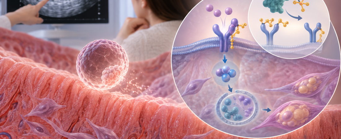
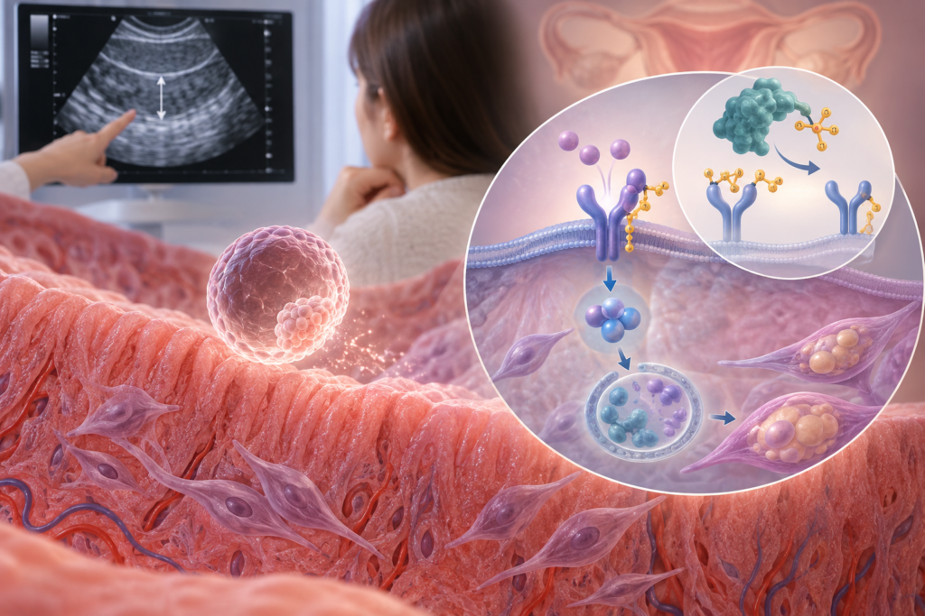
Endometrial cells possess a receptor protein called BMPR1A. Think of it as an antenna that receives “pregnancy preparation signals” arriving from outside the cell and transmits them inward.
One of the primary signaling molecules targeting this antenna is BMP2. BMP2 is an essential signaling factor governing endometrial decidualization.
The issue hinges on how effectively the antenna and signal can interact. When researchers suppressed PAPSS2, the sulfation tagging BMPR1A decreased. Remarkably, this reduction allowed BMPR1A to receive and bind BMP2 signals much more efficiently.
Put simply:
PAPSS2 decreases → Sulfate tags obstructing the antenna decrease → Pregnancy-preparation signals are transmitted more effectively.
Once the signal was received, downstream transducers known as SMAD1/5/9 were mobilized within the cell. These act as intracellular messengers relaying the external message deep into the cell’s nucleus.
Finally, another crucial cellular event occurred: autophagy.
While “autophagy” (self-eating) may sound alarming, it is actually a vital cellular housekeeping system that cleans out and recycles worn-out, unnecessary components inside the cell—much like doing a thorough housecleaning before welcoming a guest.
In this study, the activation of this internal cellular cleaning system accelerated the decidualization of endometrial stromal cells. In cellular assays, the endometrium’s capacity to accommodate the invasion of trophoblast cells—which later form the placenta—was also significantly enhanced.
What Cannot Be Known from ’10mm’ Alone
“Why did implantation fail when the endometrium was 10 mm?”
This study does not pinpoint the specific cause of implantation failure for an individual patient. However, it demonstrates at the molecular level why the simplistic equation of “thick endometrium = perfectly receptive endometrium” does not necessarily hold.
An ultrasound can visualize the thickness and shape of the endometrium. However, it cannot reveal whether the cells within are responding adequately to progesterone, whether they have sufficiently differentiated into pregnancy-competent cells, or whether they are primed to exchange biochemical crosstalk with an embryo.
Put simply, if endometrial thickness represents the “outer appearance,” decidualization represents its “functional competence.”
This does not mean endometrial thickness is unimportant. The correlation between a thin endometrium and adverse IVF outcomes remains a critical clinical concern. What this study highlights is that there is a biological domain in endometrial evaluation that cannot be captured by millimeters alone.
Should patients now undergo a PAPSS2 test? The answer is: not yet.
This study does not recommend that women experiencing implantation failure seek PAPSS2 screening. Nor is it a clinical trial proving that treatments lowering PAPSS2 improve pregnancy or live birth rates.
The researchers traced this pathway using human endometrial tissue biopsies, cultured endometrial stromal cells, transcriptomic datasets, and artificial murine decidualization models. The authors themselves emphasized that larger-scale studies are required to confirm the clinical significance of this mechanism in human reproduction.
Thus, at present, recurrent implantation failure cannot be reduced to PAPSS2 alone, nor has this technology reached the stage of being an approved diagnostic test or clinical therapy.
Still, this research is compelling for a fundamental reason. In IVF, attention has historically been overwhelmingly directed at the embryo: counting cell numbers, assigning morphological grades, tracking blastocyst progression, and verifying chromosomal euploidy.
Yet implantation is never a solo endeavor by the embryo. No matter how pristine an embryo arrives, if the endometrium has not switched into ‘pregnancy mode,’ the outcome can change entirely. In the future of endometrial research, the pivotal question may take a leap forward from “how many millimeters has it grown?” to:
“Is this endometrium truly prepared to receive an embryo?”
This study has uncovered an essential new puzzle piece explaining that invisible “functional capability” of the endometrium.
※ Study Source: Published online July 2026 in the international journal Human Reproduction. Conducted by researchers from the Centre of Reproductive Medicine, Shengjing Hospital of China Medical University. Title: “PAPSS2-mediated BMPR1A sulfation in stromal cells regulating endometrial decidualization via autophagy.” Published in Human Reproduction, Volume 41, Issue 9, Pages 1552–1569. DOI: 10.1093/humrep/deag103, PMID: 42421304.
※ The contents of this article do not replace individualized medical diagnosis or treatment. This study represents mechanistic research uncovering a novel molecular pathway regulating endometrial decidualization; it does not confirm PAPSS2 as a definitive cause of recurrent implantation failure, nor can it be immediately translated into diagnostic assays or targeted treatments. Endometrial thickness remains one of several clinical parameters evaluating implantation potential, and decisions regarding embryo transfers and clinical interventions should be determined collaboratively with an attending physician, synthesizing embryonic quality, uterine cavity health, hormonal status, and individual clinical history.
※ The images used in this article were generated using artificial intelligence (ChatGPT, OpenAI) as illustrative reference materials and do not depict real individuals.
※ Copyright © 2026 THE FERTILITY NOTES. All rights reserved. Unauthorized reproduction, duplication, distribution, scraping, AI training, or data utilization of all content, including articles, photos, and images, is strictly prohibited and subject to legal liability.