
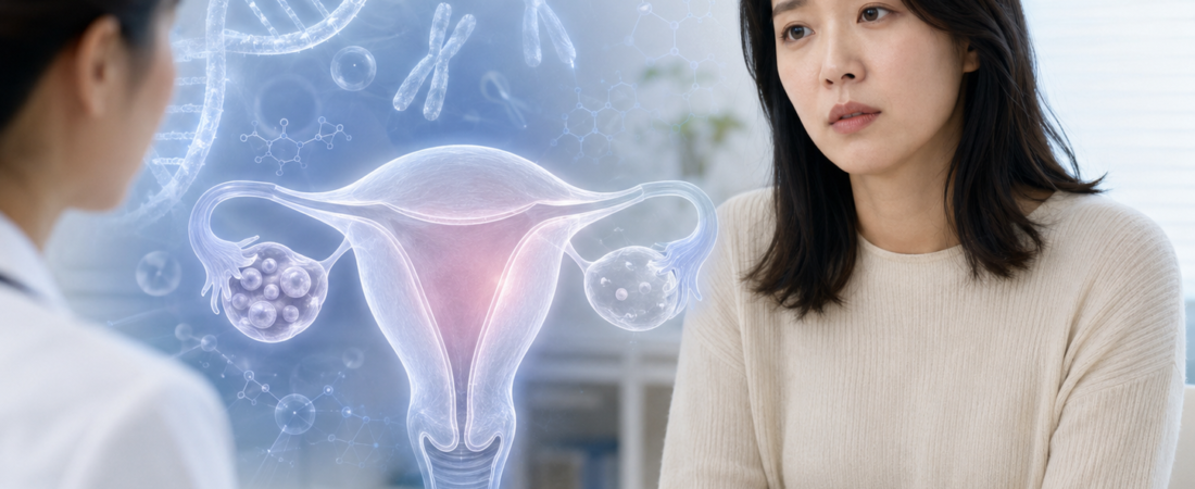
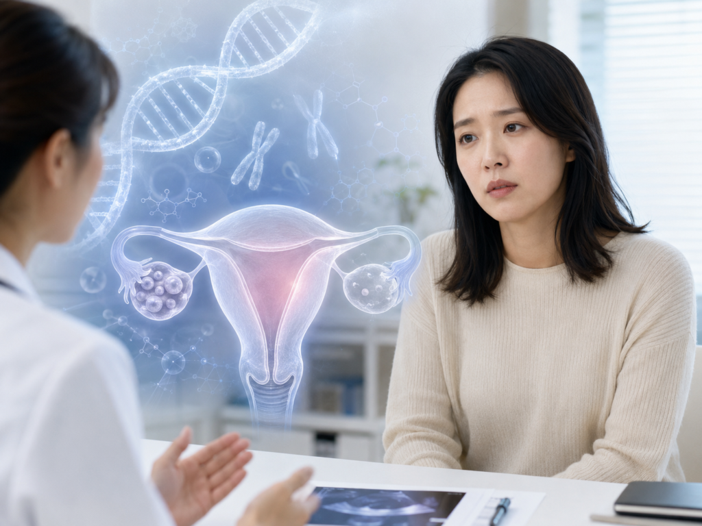
Is a Low AMH Merely ‘Premature Ovarian Aging’?… 272 Genes Uncovered Behind Primary Ovarian Insufficiency
- Comprehensive 2026 genetics review catalogs 272 genes reported across two or more unrelated POI patients
- Involvement across diverse cellular pathways: DNA repair, meiosis, and folliculogenesis; international guidelines expand clinical genetic panels
- “Do not diagnose POI by AMH alone”: Persistent menstrual irregularities and elevated FSH (> 25 IU/L) before age 40 remain essential diagnostic prerequisites
When a woman in her 30s receives an unexpectedly low Anti-Müllerian Hormone (AMH) reading during a fertility workup, she is often told that her “ovarian age is advanced.” AMH remains a valuable clinical biomarker for estimating the remaining follicular pool—the ovarian reserve.
Yet, when ovarian function declines prematurely before the age of 40, leading to irregular or absent cycles, framing the condition simply as “accelerated ovarian aging” falls short. Emerging genetic research makes it increasingly clear that Primary Ovarian Insufficiency (POI) is far more biologically complex.
A comprehensive genetics review published online in Seminars in Reproductive Medicine (March 2026) cataloged 272 distinct genes identified in two or more unrelated patients clinically diagnosed with POI. Moving past the era when the condition was blamed on just a handful of isolated mutations, science is revealing POI to be an extraordinarily heterogeneous disorder driven by interconnected cellular and molecular networks.
This does not imply that all 272 genes are definitive, causative drivers in every case. The review broadly compiled candidate genes documented in at least two unrelated individuals, meaning the underlying level of clinical evidence and penetrance varies across specific variants.
Genes at Work Across Every Stage of Oogenesis
Why are so many distinct genes linked to POI?
An oocyte is not an inert cell waiting passively inside the ovary until ovulation. From fetal life onward, female germ cell development undergoes an intricate sequence: primordial germ cells proliferate, enter meiosis, assemble into primordial follicles, and must be preserved and maintained within the ovarian cortex for decades.
Throughout this timeline, the ovary relies on an array of specialized genetic programs:
- DNA Repair and Meiotic Recombination: Genes such as MCM8, MCM9, MSH4, MSH5, and STAG3 coordinate double-strand break repair, homologous pairing, and accurate chromosome segregation during meiosis.
- Folliculogenesis and Oocyte Maturation: Transcription factors like NOBOX, FIGLA, and SOHLH1 control the formation of primordial follicles and direct early egg development.
- Cell-to-Cell Paracrine Signaling: Factors such as BMP15 and GDF9 regulate vital communication loops between the developing oocyte and surrounding somatic granulosa cells.
When a pathogenic disruption occurs in any of these pathways, the consequences are profound: the initial pool of primordial follicles formed in utero may be severely truncated, or follicles may undergo accelerated apoptotic clearance later in life.
Among established genetic causes, the premutation of the FMR1 gene (associated with Fragile X-associated primary ovarian insufficiency, or FXPOI) remains one of the most prominent single-gene drivers, alongside X-chromosome structural alterations and Turner syndrome (45,X).
Low AMH Does Not Automatically Equal POI
A critical clinical distinction must be drawn between diminished ovarian reserve (DOR) and true primary ovarian insufficiency.
While a low AMH level suggests that the numerical count of remaining primordial follicles is small, AMH alone does not establish a diagnosis of POI.
Joint clinical guidelines issued by the European Society of Human Reproduction and Embryology (ESHRE), the American Society for Reproductive Medicine (ASRM), and the International Menopause Society (IMS) define POI as the premature cessation or severe decline of ovarian activity before age 40.
Standard diagnostic criteria require:
- Menstrual Disturbance: Amenorrhea or oligomenorrhea lasting for at least 4 consecutive months.
- Elevated Gonadotropins: Serum Follicle-Stimulating Hormone (FSH) exceeding 25 IU/L on two occasions (measured 4 to 6 weeks apart if clinical confirmation is required).
The consensus guidelines explicitly state that AMH should not be used as a first-line diagnostic test for POI. While AMH can offer supportive prognostic context when FSH results are borderline, clinical evidence remains insufficient to diagnose current POI or reliably forecast future POI solely on the basis of an isolated AMH value.
A woman in her 30s with a low AMH who continues to have regular ovulatory cycles should not be told that her ovaries have “stopped functioning.”
Moving Beyond “Idiopathic”: Genetic Panels Step Forward
For women diagnosed with POI, identifying the underlying etiology has historically been difficult, with the majority labeled “unexplained” or “idiopathic.” That approach is shifting.
Current international guidelines recommend that women presenting with POI without an obvious iatrogenic cause (such as prior gonadotoxic chemotherapy, pelvic radiation, or extensive bilateral ovarian surgery) undergo:
- Karyotyping: To screen for numerical or structural chromosomal anomalies.
- FMR1 Premutation Testing: To evaluate CGG repeat expansions.
Where resources and accredited genetic counseling are available, guidelines support extending evaluations to multi-gene Next-Generation Sequencing (NGS) panels. Importantly, experts emphasize that patients should not be excluded from genetic workups based purely on chronological age at diagnosis.
Pinpointing a genetic etiology does far more than provide emotional closure regarding the cause of subfertility. Discovering a specific pathogenic variant allows clinicians to evaluate reproductive risks for biological sisters and daughters, while also screening for associated extra-ovarian health issues—such as autoimmune endocrinopathies, sensory deficits, or metabolic disorders.
When a genetic explanation is absent, autoimmune screening becomes essential. Clinical guidelines recommend testing for 21-hydroxylase autoantibodies (21-OH Ab) to assess adrenal autoimmunity, alongside comprehensive thyroid autoantibody and functional evaluations.
POI Does Not Mean the Ovary Is Permanently ‘Dead’
Because of the historical name “premature ovarian failure,” many patients assume the ovary is completely and irreversibly dormant. That assumption is not always accurate.
In non-surgical, spontaneous POI, unpredictable intermittent ovarian activity can occur, with spontaneous ovulation and natural conception documented in roughly 5% of cases. For this reason, the medical community prefers the term ovarian “insufficiency” over “failure.”
Nevertheless, because the chances of spontaneous conception are low and no medical treatment has been proven to reliably restore follicle activation, oocyte donation remains the established, evidence-based assisted reproductive pathway for achieving pregnancy.
Furthermore, POI carries health implications that extend well beyond fertility. Sustained estrogen deficiency in a young woman accelerates bone mineral density loss and elevates long-term cardiovascular and metabolic risks. In the absence of strict contraindications, clinical guidelines strongly recommend initiating Hormone Replacement Therapy (HRT), continuing treatment until the average physiological age of natural menopause (approximately 50 to 51 years).
Reconceptualizing the Disease
The takeaway from recent genetic advances is clear: when a woman under 40 experiences a premature loss of ovarian function, it should not simply be brushed off as “unlucky early aging.”
Normal oogenesis and lifelong follicle maintenance depend on hundreds of coordinated genes. Rather than a single disease entity, POI represents a broad, complex spectrum where varied molecular breakdowns—spanning DNA repair, meiotic checkpoint controls, and follicular signaling—converge on the shared clinical endpoint of premature ovarian decline.
As genomic sequencing deepens, the veil is lifting on what was once filed away as “unexplained.”
Medical Source & Study Reference
- Journal: Seminars in Reproductive Medicine, Volume 43, Issue 4, pp. 296–322 (Published online March 2, 2026).
- Review Title: Genetics of Primary Ovarian Insufficiency
- Authors: Yatsenko SA, Rajkovic A
- DOI: 10.1055/a-2806-2597 | PMID: 41771295
※ This article was synthesized based on the state-of-the-art genetics review published in Seminars in Reproductive Medicine (2026) alongside international clinical consensus guidelines from ESHRE, ASRM, and the IMS. It does not replace individualized clinical diagnosis or medical care, and specific medical decisions should always be made in consultation with a qualified reproductive endocrinologist or medical geneticist.
※ The images associated with this article were generated using generative AI (ChatGPT, OpenAI) as illustrative visual references and do not depict real individuals.