
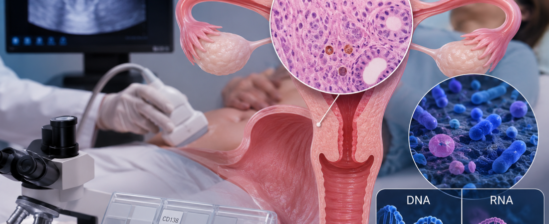
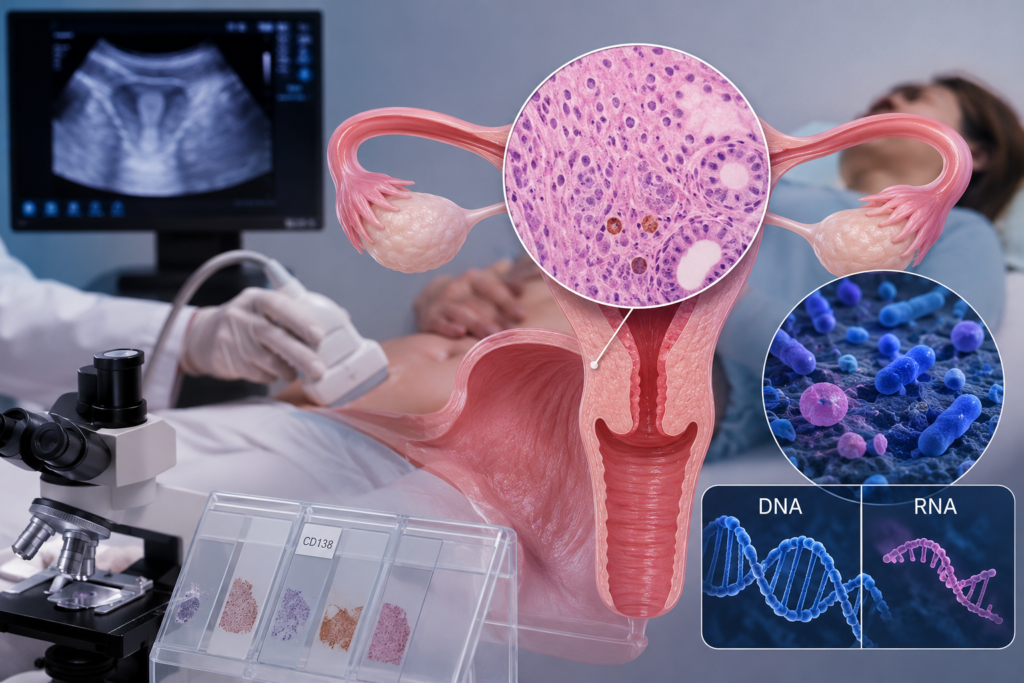
- 160 Women Tested Ahead of First Frozen Embryo Transfer: Chronic endometritis confirmed in 18.1%
- CD138 Immunohistochemistry, Histopathology, and Molecular Assays Used: Difficult to uncover through ultrasound alone
- Looking Beyond DNA to RNA Profiling: Demonstrating the potential to distinguish ‘actively functioning microorganisms’
For women undergoing fertility treatment, hearing the ultrasound verdict—”Your uterine lining looks clean and has good thickness”—brings an immediate sense of relief. However, the fact that the endometrial cavity appears normal on an ultrasound screen does not necessarily mean the implantation environment itself is healthy.
In a prospective study presented at the 2026 Annual Meeting of the European Society of Human Reproduction and Embryology (ESHRE), researchers performed comprehensive testing on women preparing for their first frozen embryo transfer (FET) who showed no structural abnormalities on transvaginal ultrasound. The result: chronic endometritis (CE) was discovered in 18.1% of patients—nearly one in every five women.
Chronic endometritis is a persistent, low-grade inflammatory state of the endometrial lining. Unlike acute endometritis, it rarely presents with obvious clinical symptoms such as fever, severe pelvic pain, or foul-smelling discharge, meaning it is often detected purely by chance during subfertility evaluations. While its link to recurrent implantation failure and repeated pregnancy loss has been studied for years, the lack of unified international diagnostic criteria has remained an ongoing challenge in clinical practice.
Identified in 29 Out of 160 Women with Normal Ultrasounds
The study evaluated 160 women scheduled for their first frozen embryo transfer between July 2022 and December 2025. Although all participants had transvaginal ultrasounds confirming a structurally normal endometrial cavity, they underwent additional diagnostic hysteroscopy and endometrial biopsies 1 to 4 months prior to embryo transfer.
The researchers combined standard histopathological evaluation with CD138 immunohistochemical (IHC) staining to detect stromal plasma cells, alongside real-time polymerase chain reaction (RT-PCR) to test for bacterial genetic material. In total, 29 out of the 160 women (18.1%) were diagnosed with chronic endometritis across at least one of these diagnostic modalities.
Molecular testing identified Mycoplasma and Ureaplasma in 10 cases each, with Escherichia coli also detected; several patients exhibited co-infections with multiple organisms. In other cases, abnormal findings were captured solely through CD138 staining or standard histopathology.
These findings highlight a key clinical reality: transvaginal ultrasound is designed to inspect macroscopic uterine anatomy—such as endometrial thickness, polyps, and fibroids—not microscopic inflammation or subtle shifts in local immune cells. The hallmark of chronic endometritis, plasma cell infiltration into the endometrial stroma, can only be diagnosed by directly examining tissue.
After Antibiotic Treatment, the Pregnancy Rate Gap Disappeared
The most encouraging data emerged when women diagnosed with chronic endometritis underwent targeted antibiotic therapy prior to frozen embryo transfer.
Among treated patients with chronic endometritis, the clinical pregnancy rate reached 55.2%, compared to 62.6% in the disease-free control group. The ongoing pregnancy rates were 44.8% and 48.1%, respectively. There was no statistically significant difference between the two groups. These results suggest that identifying and treating chronic endometritis can help restore clinical reproductive outcomes for affected patients.
However, these findings should not be interpreted to mean that every IVF patient requires routine endometrial biopsies or preemptive antibiotic courses.
This was a single-center study, and the total number of confirmed chronic endometritis cases was relatively small at 29 patients. Furthermore, comprehensive molecular PCR and CD138 staining were not applied identically across all 160 women; rather, reflex testing was performed primarily on patients who demonstrated suspicious vascular or mucosal signs during diagnostic hysteroscopy—a limitation acknowledged by the authors. In addition, standardized diagnostic thresholds for chronic endometritis continue to vary across different clinics worldwide.
Does Detecting DNA Mean the Bacteria Are ‘Alive’?
Alongside tissue biopsies, a deeper biological question has recently gained traction:
If bacterial DNA is detected in the endometrial cavity, does that prove the microorganisms are actually alive and proliferating?
Conventional microbial assays rely heavily on DNA sequencing. The issue is that DNA is a stable molecule that can be detected from non-viable bacteria, dead bacterial fragments, or cell-free genetic debris. Therefore, a positive DNA signal alone cannot definitively prove that a specific microbe is actively colonizing and exerting pathogenic effects within the endometrium.
To address this, researchers are turning toward metatranscriptomic profiling (RNA analysis). Because RNA is produced during active gene expression and ongoing metabolic processes, it provides a clearer picture of which microorganisms are functionally active in real time. A study published in Human Reproduction Open in 2026 confirmed that bacterial community profiles can look substantially different when analyzed through DNA-based sequencing compared to RNA-based transcriptomics.
Ureaplasma, Gardnerella, and Mycoplasma Captured on RNA
A companion study presented at ESHRE 2026 evaluated this distinction directly, performing simultaneous DNA and RNA sequencing on endometrial biopsy tissue from 68 women with chronic endometritis.
Initial findings revealed that concordance between DNA and RNA detection was surprisingly low. Notably, organisms such as Ureaplasma, Gardnerella, and Mycoplasma were significantly more prominent and transcriptionally active on RNA profiling.
This demonstrates that reproductive microbiome research is shifting away from merely asking “Is bacterial DNA present?” toward determining “Which microorganisms are actively functioning in this environment?”
Nevertheless, this remains an ongoing preliminary trial. The authors noted that integrated profiling of local immune cell populations and host endometrial gene expression is still underway, and final conclusive data have yet to be published. It is therefore too early to assert that specific bacteria are the direct, isolated cause of chronic endometritis, or that routine clinical RNA sequencing can single-handedly diagnose the root cause of implantation failure today.
From ‘Embryo-Centric’ Care to the Endometrial Microenvironment
The broader takeaway from these studies is clear:
When patients experience repeated implantation failure despite transferring high-quality embryos, the investigation must look beyond embryonic chromosomes and visual grading to examine the inflammatory, immune, and microbial microenvironment of the receptive endometrium.
Chronic endometritis can exist silently in women whose ultrasounds appear entirely normal and who report no physical symptoms. At the same time, finding trace microbial DNA should not serve as an automatic green light for indiscriminate antibiotic overuse.
The ongoing challenge for reproductive medicine is to look beyond binary questions of “presence or absence” to uncover which microorganisms are genuinely active, how they alter the endometrial immune response, and whether those molecular shifts ultimately dictate implantation and live birth outcomes.
The hidden world of the endometrium—invisible on an ultrasound monitor—is establishing itself as an essential frontier in modern fertility care.
※ This article was written based on research presented at the 2026 Annual Meeting of the European Society of Human Reproduction and Embryology (ESHRE) and published in the July 2026 Supplement of Human Reproduction (Volume 41, Supplement 1): “Prospective study on prevalence, associated factors and reproductive outcomes of chronic endometritis before first elective frozen embryo transfer” (Presentation L26/P-322, Abstract deag083.658, DOI: 10.1093/humrep/deag083.658) and “Human and microbial multi-omic landscape of chronic endometritis” (Presentation L26/P-343, Abstract deag083.679, DOI: 10.1093/humrep/deag083.679). Methodological comparisons between DNA and RNA microbial sequencing also referenced “16S rRNA gene sequencing and metatranscriptomics of the female genital tract microbiome” published in Human Reproduction Open (Issue 1, 2026, Article hoag001, DOI: 10.1093/hropen/hoag001). It does not replace individualized clinical diagnosis or medical care, and specific treatment decisions should always be made in consultation with a qualified reproductive specialist.
※ Image: AI generated (ChatGPT, OpenAI) / For illustrative reference only.