
- Analyzing lesions, the eutopic endometrium, and menstrual blood: Mapping the direct biological link between chronic inflammation and progressive fibrosis
- Identifying CHI3L1+ fibroblasts and immune aberrations: Scientific weight shifts toward a “whole-uterine microenvironmental disease”
- Inflammatory signatures confirmed in menstrual effluent: Opening new doors for non-invasive diagnostics and targeted anti-fibrotic therapies
The way we understand uterine adenomyosis is undergoing a fundamental shift. For decades, the condition was defined almost exclusively through an anatomical lens: endometrial tissue lining the inner uterine cavity abnormally invading the muscular myometrial wall.
Recent scientific findings, however, increasingly challenge this simplistic view. Rather than a mere “spatial displacement of tissue,” adenomyosis is emerging as a systemic uterine disorder characterized by persistent chronic inflammation and progressive tissue fibrosis.
A single-cell profiling study presented at the 2026 Annual Meeting of the European Society of Human Reproduction and Embryology (ESHRE) brought this concept into sharp focus. By analyzing not only ectopic myometrial lesions but also the normally situated endometrium and shed menstrual blood, researchers mapped these cellular disturbances as an interconnected disease environment. The presentation earned the prestigious Basic Science Award for oral presentation at ESHRE 2026.
The research team evaluated a total of 145 clinical specimens collected from 80 women with adenomyosis and 65 disease-free controls. Subsets of these samples underwent single-cell RNA sequencing (scRNA-seq) alongside comprehensive cytokine profiling across the myometrium, endometrium, and menstrual effluent.
Unlike traditional bulk sequencing methods that average signals across tens of thousands of mixed cells, single-cell analysis dissects the precise gene-expression programs of individual cells one by one.
The Discovery of “CHI3L1+ Fibroblasts” in Adenomyosis
The most striking cellular hallmark uncovered in the adenomyotic lesions was a distinct population of CHI3L1-positive (CHI3L1+) fibroblasts.
Fibroblasts are responsible for synthesizing the extracellular matrix that structurally supports tissues and coordinates wound healing. When aberrantly or chronically overactivated, however, they produce excess collagen and matrix proteins, driving pathological tissue stiffening and fibrosis.
The study demonstrated that adenomyotic lesions harbor a specialized subpopulation of fibroblasts marked by robust expression of CHI3L1 (Chitinase-3-like protein 1). Concurrently, the researchers observed a marked accumulation of exhausted CD8+ T cells alongside pathologically elevated concentrations of pro-inflammatory and pro-fibrotic cytokines—including Interleukin-2 (IL-2), IL-4, IL-5, IL-16, Interferon-gamma (IFN-gamma), Transforming Growth Factor-beta1 (TGF-beta1), and basic Fibroblast Growth Factor (bFGF). The authors characterized this niche as an active storm where chronic inflammation, tissue fibrosis, neurogenesis, and hyper-vascularization unfold simultaneously.
The biological significance of these CHI3L1+ fibroblasts aligns with recent discoveries. A 2025 study published in Advanced Science demonstrated that calcitonin gene-related peptide (CGRP), released by local sensory nerve fibers transmitting pelvic pain, can directly reprogram resting fibroblasts into CHI3L1-hypersecreting phenotypes, accelerating myometrial fibrosis. This suggests that the severe pain and structural fibrosis characteristic of adenomyosis are not separate clinical symptoms, but rather two sides of a shared neuro-fibrotic feedback loop.
More Than an Isolated Lesion: The Eutopic Endometrium Changes Too
Crucially, the cellular abnormalities were not restricted to the lesions buried deep within the muscular myometrial wall.
Within the eutopic endometrium—the normal inner lining of the uterus where an embryo must implant—macrophages and stromal cells exhibited pronounced pathological alterations. Cellular stress-response markers, such as heat shock proteins (HSPs), were significantly elevated, accompanied by the upregulation of core fibrotic and extracellular matrix-remodeling genes, including MRC1, CD163, SERPINE1, CCN2, THBS1, and COL4A1.
This demonstrates why adenomyosis cannot be viewed simply as “stray endometrial tissue in the muscle layer.” The receptive endometrium itself—the biological soil where an embryo must attach—is fundamentally altered by widespread inflammatory and fibrotic cascades.
In reproductive medicine and fertility care, this distinction is paramount. Even when transferring a chromosomally normal, top-tier blastocyst, altered endometrial receptivity can derail the biochemical dialogue required for successful implantation.
The researchers noted that altered bFGF expression within the eutopic endometrium may offer a functional molecular clue explaining the elevated rates of recurrent implantation failure (RIF) seen in patients with adenomyosis, though direct interventional proof warrants ongoing study.
Could Menstrual Blood Become a “Liquid Biopsy” for the Uterus?
Another compelling horizon illuminated by the study lies in menstrual blood.
Currently, diagnosing adenomyosis depends primarily on transvaginal ultrasound (TVUS) or pelvic Magnetic Resonance Imaging (MRI). However, detecting diffuse, subtle, or early-stage lesions remains challenging and heavily reliant on operator expertise.
The research team discovered that the distinct inflammatory and immune signatures identified inside the uterus were clearly reflected within menstrual blood. Cellular shifts and local cytokine surges occurring deep inside the uterine cavity leave measurable molecular traces in the shed effluent, suggesting that menstrual blood profiling could eventually evolve into a non-invasive diagnostic tool.
This does not mean a routine “single-drop menstrual blood test” is ready for immediate clinic adoption today. The sample cohort evaluated via single-cell sequencing and cytokine panels was relatively small, and developing a validated diagnostic assay requires standardized multi-center trials to pinpoint exact clinical biomarker thresholds.
Nevertheless, the biological premise is profound. Menstrual blood directly captures shed endometrial cells, tissue fragments, secretions, and immune regulators. Analyzing this fluid with high-resolution molecular tools offers the potential to read real-time uterine pathology without requiring invasive tissue biopsies.
Expanding the Therapeutic Horizon
The core takeaway of this research is unambiguous: defining adenomyosis merely as the mechanical invasion of endometrial glands into the muscle is no longer sufficient.
It is a dynamic, organ-wide condition where chronic inflammation persists, immune cell equilibrium breaks down, and activated fibroblasts progressively fibrose the uterine architecture—alterations that extend from focal myometrial lesions to the receptive endometrium and out into menstrual fluid.
If this paradigm continues to solidify, future therapies will likely expand beyond traditional approaches that focus solely on suppressing ovarian hormones or surgically reducing lesions. The frontier of adenomyosis management is moving toward targeted interventions that modulate chronic inflammation, halt tissue fibrosis, and restore the biological health of the endometrium for women preparing for pregnancy.
Medical Source & Study Information
- Conference: European Society of Human Reproduction and Embryology (ESHRE) 42nd Annual Meeting (2026)
- Journal Publication: Human Reproduction, Volume 41, Supplement 1 (July 2026 Issue)
- Study Title: Systemic inflammatory and fibrotic landscape across lesions, endometrium, and menstrual blood in adenomyosis: an integrated single-cell RNA sequencing and cytokine analysis
- Abstract Number: L26/O-078 | Paper Number: deag083.078 | DOI: 10.1093/humrep/deag083.078
- Recognition: Awarded the ESHRE 2026 Basic Science Award for Oral Presentation
※ This article was synthesized based on the basic science study presented at the 2026 ESHRE Annual Meeting and published in the Human Reproduction supplement, alongside literature in gynecological pathology and single-cell transcriptomics. It does not replace individualized clinical diagnosis or medical care, and specific treatment decisions should always be made in consultation with a qualified reproductive endocrinologist or gynecologist.
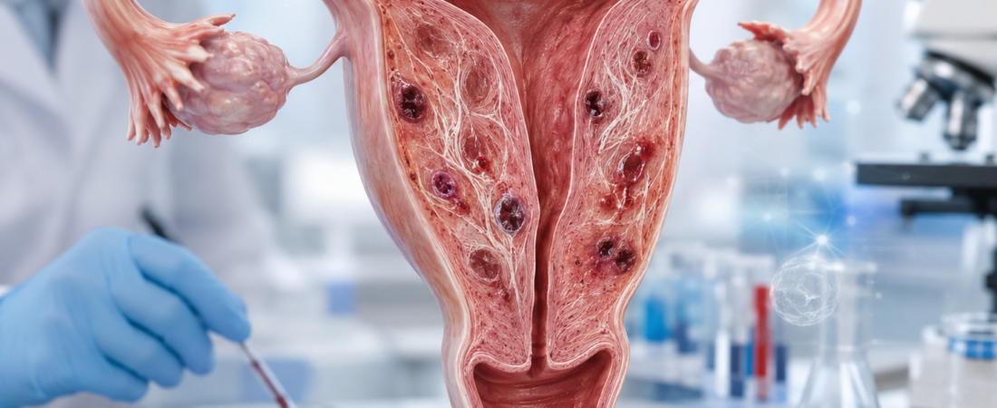
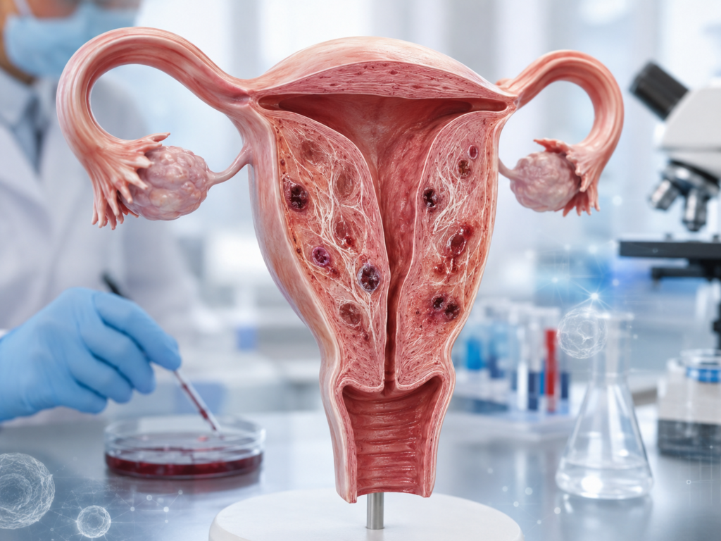
※ The images associated with this article were generated using generative AI (ChatGPT, OpenAI) as illustrative visual references and do not depict real individuals.