
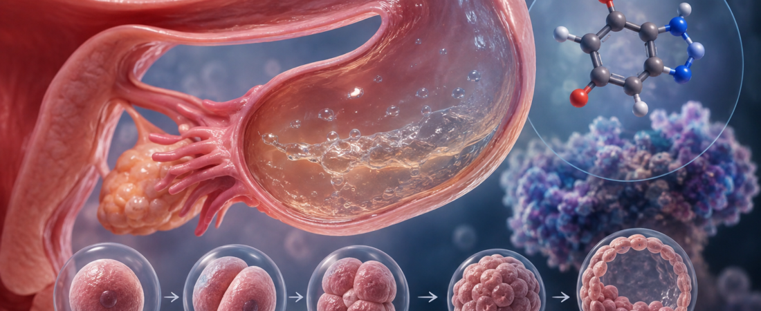
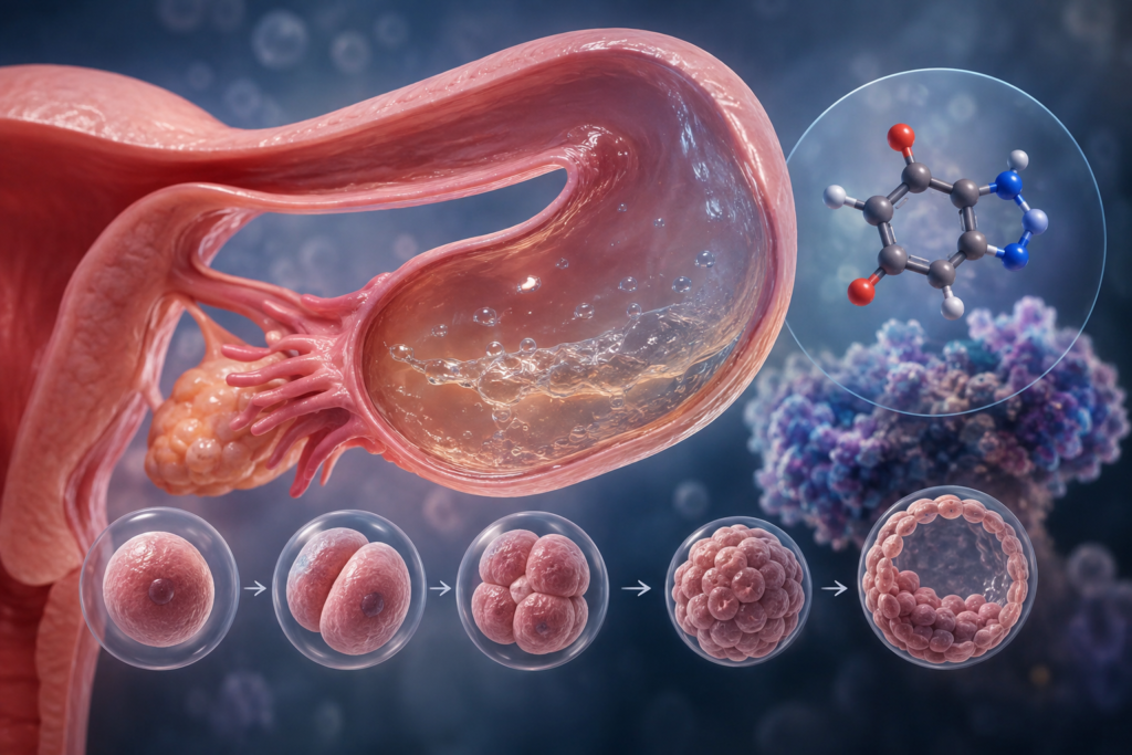
- Tubal Fluid Analysis Across 49 Hydrosalpinx Patients and 52 Controls: Confirmed purine metabolic dysregulation and marked inosine accumulation
- Mouse Embryos Arrest at the 2- to 4-Cell Stage: Inosine impairs the critical maternal-to-zygotic transition (MZT) and embryonic genome activation
- Reframing the Fallopian Tube: Moving beyond a mechanical conduit to the embryo’s very first active biochemical microenvironment
When a patient preparing for In Vitro Fertilization (IVF) is diagnosed with a hydrosalpinx (a fluid-filled, obstructed fallopian tube), clinicians frequently recommend laparoscopic salpingectomy or proximal tubal occlusion prior to embryo transfer. The prevailing clinical rationale has long focused on the mechanical backflow of toxic tubal fluid into the endometrial cavity, which physically flushes out embryos or impairs endometrial receptivity.
However, emerging research reveals that the reduced success rates associated with hydrosalpinx may stem from more than mechanical regurgitation alone. A specific metabolite accumulated within hydrosalpinx fluid has been shown to directly arrest early embryonic division.
In a study published online in eBioMedicine (and in its June 2026 issue), a research team from Peking University Third Hospital reported that inosine—a purine metabolite—was significantly elevated in the tubal fluid of patients with hydrosalpinx and directly correlated with early embryonic developmental arrest.
Beyond “Stagnant Fluid”: Identifying Purine Dysregulation
A hydrosalpinx develops when the distal fimbrial end of the fallopian tube becomes occluded following pelvic inflammatory disease (PID), pelvic surgery, or endometriosis, leading to distension and fluid accumulation.
In reproductive medicine, hydrosalpinx is a major prognostic factor. Meta-analyses cited by the American Society for Reproductive Medicine (ASRM) demonstrate that an untreated hydrosalpinx can reduce implantation, clinical pregnancy, and live birth rates by approximately 50%. Proposed mechanisms historically included mechanical wash-out of embryos, altered endometrial integrin expression, and general embryotoxicity of the fluid.
The Peking University research team investigated the exact biochemical composition causing this toxicity.
Utilizing untargeted metabolomic profiling, the researchers compared tubal fluid samples collected from 49 women with hydrosalpinx against 52 control patients. The analysis revealed that purine metabolism was among the most heavily dysregulated pathways in the hydrosalpinx cohort, marked by a pronounced accumulation of inosine.
In Vitro Functional Assays: Embryos Arrest at the 2- to 4-Cell Stage
To evaluate the biological impact of inosine, the researchers conducted dose-response functional assays using mouse embryos:
- High Concentrations (25 μM and 50 μM): Culturing zygotes in inosine-supplemented media caused extensive developmental arrest at the 2-cell to 4-cell stage.
- Lower Concentration (12.5 μM): Significantly reduced the blastocyst formation rate. Embryos that managed to reach the blastocyst stage exhibited lower total cell counts and impaired inner cell mass (ICM) proliferation.
- Irreversible Damage: Embryos exposed to inosine during the earliest cleavage stages failed to fully recover developmental potential even after being washed and transferred into standard, inosine-free culture medium.
These findings suggest that a hydrosalpinx environment exerts embryotoxic effects from the moment of fertilization, disrupting cleavage divisions long before implantation in the uterus.
Mechanism: Disrupting the Maternal-to-Zygotic Transition (MZT)
Transcriptomic and translatomic analyses elucidated the molecular mechanism underlying this arrest.
Immediately following fertilization, an embryo initially relies on maternal transcripts and proteins stored within the ooplasm. At a defined developmental milestone (the 2-cell stage in mice, and the 4- to 8-cell stage in humans), the embryo must degrade maternal factors and switch on its own genomic machinery—a process known as the Maternal-to-Zygotic Transition (MZT) or Embryonic Genome Activation (EGA).
Inosine exposure directly disrupted this critical transition:
- Overall protein translation efficiency was significantly suppressed.
- Cytoskeletal architecture and spindle organization were impaired.
- Embryonic genome activation failed to initiate properly, halting the developmental cascade required to progress past early cleavage stages.
Rescuing Development via PNP Inhibition
The research team also examined Purine Nucleoside Phosphorylase (PNP), a key enzyme involved in inosine metabolism.
When researchers pharmacologically inhibited PNP using forodesine or knocked down its expression genetically, the inosine-induced developmental arrest was partially rescued, allowing a subset of arrested embryos to resume cleavage and development.
This indicates that inosine accumulation is a functional driver of developmental failure rather than an innocent metabolic byproduct. Furthermore, it highlights the PNP metabolic pathway as a potential pharmacological target for future research into tubal-factor subfertility.
Study Limitations
While the human tubal fluid metabolomic profiling was conducted on clinical patient samples, the embryonic toxicity and mechanistic rescue experiments were performed in rodent models. Whether identical inosine concentration thresholds and downstream translational arrests occur in human embryos requires further verification.
Clinical Implications: Reframing Fallopian Tube Biology
These findings provide a deeper biochemical foundation for existing clinical practice:
- Active Reproductive Organ: The fallopian tube is not a passive anatomical pipe; it is the active biological environment where fertilization and the earliest cell divisions unfold.
- Biochemical Rationale for Surgical Management: Current guidelines from the World Health Organization (WHO) and ASRM recommend prophylactic salpingectomy or proximal tubal occlusion for hydrosalpinges prior to IVF. This study provides concrete metabolic evidence—beyond simple hydrostatic fluid reflux—explaining why isolating the uterine cavity from toxic tubal metabolites is biologically essential to safeguard embryonic development.
Medical Source & Study Information
- Authors: Research team at Peking University Third Hospital
- Journal: eBioMedicine (Part of The Lancet Discovery Science), Published online May 21, 2026; June 2026 Issue.
※ This article was synthesized based on research from Peking University Third Hospital published in eBioMedicine (2026) alongside clinical guidelines from the ASRM and WHO. It does not replace individual clinical diagnosis or medical care, and specific treatment decisions should always be made in consultation with a qualified reproductive endocrinologist.
※ The images associated with this article were generated using generative AI (ChatGPT, OpenAI) as illustrative visual references and do not depict real individuals.