
“Infusing PRP into Thin Endometrium Thickened It by 0.95 mm… But Childbirth Rates Did Not Rise”
- Analysis of 280 Embryo Transfers Across Spanish and Italian Fertility Centers: Endometrial thickness gain averaged 0.95 mm in the PRP group vs. 0.16 mm in controls
- No Statistically Significant Improvement Across Biochemical, Clinical, Ongoing Pregnancy, or Live Birth… Applying the brakes to the “Thickness = Implantation Capacity” dogma
- Multivariate Adjusted Analysis Confirms No Significant Association with Live Birth: Embryo-related factors and oocyte origin exerted a far greater influence on success
“Your uterine lining is too thin; we need to postpone the transfer again.”
For patients undergoing In Vitro Fertilization (IVF), endometrial thickness is a sensitive and stressful metric. Even when high-grade embryos are available, an insufficiently developed endometrium often forces clinicians to delay or cancel the transfer.
When standard exogenous estrogen protocols fail to thicken the endometrium past the conventional 7 mm threshold, clinics often explore adjuvant therapies—such as sildenafil (Viagra), granulocyte colony-stimulating factor (G-CSF), and autologous Platelet-Rich Plasma (PRP).
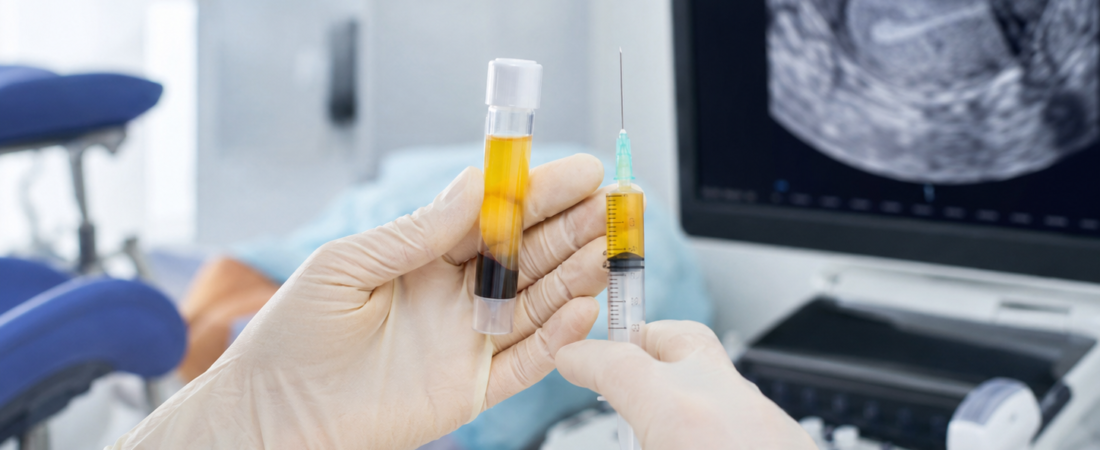
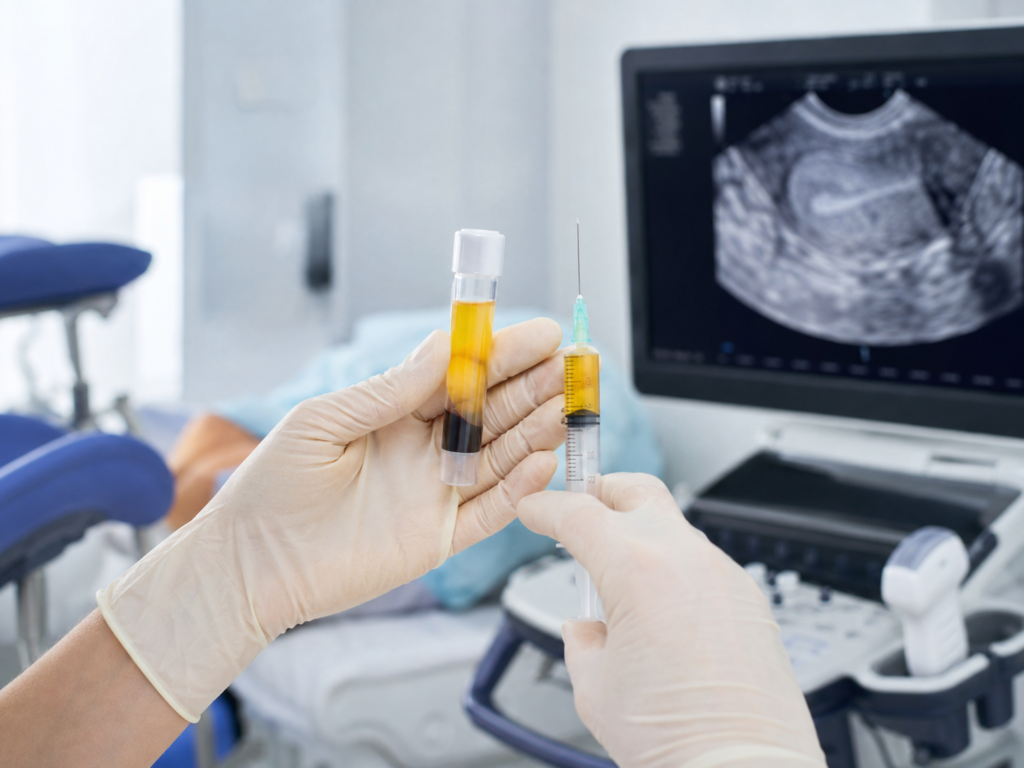
Among these, intrauterine PRP infusion has garnered significant clinical attention in recent years. The rationale relies on concentrating platelets from the patient’s own peripheral blood, delivering high concentrations of autologous growth factors and cytokines directly to the uterine cavity to promote tissue regeneration and angiogenesis.
While small observational series frequently reported that “the lining grew by 1–2 mm” and “canceled cycles were salvaged,” a critical scientific question remained unanswered: Does a thicker endometrium achieved via PRP actually translate into more live births?
The IVIRMA Multicenter Study: PRP Successfully Thickened the Lining
A multicenter study addressing this question directly was published online in Fertility and Sterility on August 5, 2026.
Researchers from the IVIRMA group and collaborating institutions analyzed 280 embryo transfers performed across fertility centers in Spain and Italy. The study cohort comprised women with refractory thin endometrium (measuring 3.0 to 6.9 mm) and a history of at least two previous implantation failures:
- PRP Group: 94 transfer cycles received intrauterine autologous PRP infusions prior to transfer.
- Control Group: 186 transfer cycles proceeded without PRP intervention.
From a purely anatomical standpoint, PRP demonstrated clear proliferative efficacy:
- Final Endometrial Thickness: Averaged 6.65±1.0 mm in the PRP group vs. 6.12±0.65 mm in the control group.
- Net Increase from Prior Cycle: The PRP group gained an average of +0.95±0.99 mm, whereas the control group increased by only +0.16±0.77 mm (P<0.05).
In short, PRP successfully improved the morphological measurements visible on ultrasound.
The Clinical Paradox: No Improvement in Implantation or Live Birth
The unexpected divergence emerged when evaluating downstream reproductive outcomes. Despite achieving a measurably thicker endometrial stripe, patients in the PRP group showed no statistically significant improvements in pregnancy or childbirth metrics.
Comparing clinical milestones between groups:
- Biochemical Pregnancy Rate: 43.62% (PRP) vs. 49.46% (Control)
- Clinical Pregnancy Rate: 35.11% (PRP) vs. 44.62% (Control)
- Live Birth Rate (LBR): 24.47% (PRP) vs. 36.76% (Control)
Because patients in the PRP cohort presented with thinner baseline endometria and poorer baseline prognostic factors, raw percentage differences should not be interpreted as evidence that PRP harmed outcomes.
However, after adjusting for confounding variables—including female age, body mass index, embryo developmental stage, and oocyte source—multivariate analysis confirmed no statistically significant association between PRP treatment and live birth:
- Adjusted Odds Ratio (aOR) for Live Birth: 0.56 (95% CI: 0.30–1.05; not statistically significant).
Instead, live birth outcomes were driven predominantly by embryo-related variables (such as oocyte origin and ploidy status) rather than whether PRP had been infused. The authors cautioned against the oversimplified assumption that “artificially increasing endometrial thickness will automatically improve pregnancy and delivery chances.”
“7 mm” Is a Diagnostic Proxy, Not the Cause of Implantation
While a thin endometrium correlates with lower baseline implantation rates, clinical medicine must distinguish correlation from causation. The observation that thin-lining cohorts experience lower average pregnancy rates does not mean that mechanically expanding the tissue by 1 mm will raise success rates by a corresponding margin.
Ultrasound-measured thickness is merely a gross anatomical surrogate marker. Successful blastocyst implantation requires:
- Synchronized molecular endometrial receptivity (the window of implantation)
- Competent embryonic-endometrial biochemical cross-talk
- Functional maternal vascular remodeling
- Embryonic chromosomal euploidy and metabolic viability
Artificially expanding a lining from 6.0 mm to 7.0 mm is biologically distinct from transforming it into a functionally receptive endometrium. The primary takeaway of this trial is that improving a surrogate ultrasound measurement does not automatically guarantee improved clinical live births.
Study Limitations and Current Evidence Base
These findings do not permanently invalidate PRP, but they contextualize its clinical role within the broader literature:
- Design Constraints: This was a retrospective cohort study rather than a randomized controlled trial (RCT), and baseline patient characteristics differed between cohorts. Furthermore, PRP preparation protocols, infusion timings, and endometrial preparation regimens varied across participating centers.
- Systematic Review Findings: A 2026 systematic review focusing on randomized trials of PRP in reproductive medicine noted that while PRP frequently improves endometrial thickness, the overall certainty of evidence regarding clinical pregnancy and live birth remains rated as “Very Low.” Crucially, only two published RCTs have reported definitive live birth outcomes, precluding robust meta-analytic pooling.
Consequently, the most evidence-based conclusion is not that “PRP has zero biological activity,” but rather that “while autologous PRP can induce modest anatomical thickening in thin endometria, robust clinical evidence confirming that it raises live birth rates remains lacking.”
Conclusion
For patients navigating refractory thin endometrium, the ultimate clinical goal is not achieving a nominal “7 mm” readout on an ultrasound screen, but sustaining a viable pregnancy to term.
This multicenter cohort study underscores that fertility adjuvants must be held to the standard of definitive live birth outcomes rather than surrogate anatomical endpoints.
Medical Source & Study Information
- Journal: Fertility and Sterility (Official Journal of the American Society for Reproductive Medicine, ASRM), Published online August 5, 2026.
- Study Title: Autologous platelet-rich plasma on endometrial growth and reproductive outcomes in women with thin endometrium: a multicentre cohort study
- Authors: IVIRMA Research Group et al.
※ This article was synthesized based on the multicenter cohort study published in Fertility and Sterility (August 2026) alongside relevant systematic reviews and clinical guidelines in reproductive medicine. It does not replace individualized clinical diagnosis or medical care, and specific treatment decisions should always be made in consultation with a qualified reproductive specialist.
※ The images associated with this article were generated using generative AI (ChatGPT, OpenAI) as illustrative visual references and do not depict real individuals.