
Focusing on Molecular Diagnostics like PTEN and PIK3CA Opening an Era of “Same Diagnosis, Different Treatment”
“Your endometrium is thick.”
This is a common phrase heard in obstetric and gynecological clinics. While most cases are temporary changes related to the menstrual cycle, if abnormal uterine bleeding persists or the endometrium remains thick even after menopause, a biopsy is often recommended. The representative disease diagnosed at this stage is endometrial hyperplasia.
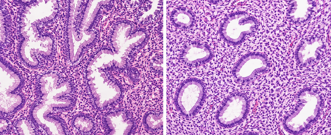
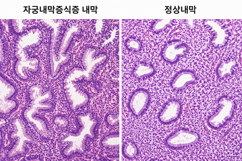
Endometrial hyperplasia refers to a state where the cells lining the inside of the uterus proliferate abnormally. It primarily occurs when there is persistent estrogen stimulation without sufficient counteracting progesterone, and common risk factors include obesity, Polycystic Ovarian Syndrome (PCOS), chronic anovulation, and diabetes.
Many women, upon receiving this diagnosis, worry first: “Does this mean I have cancer?” However, not all endometrial hyperplasia progresses to cancer.
Endometrial hyperplasia without atypical cells can often be restored to normal through hormone therapy, and the risk of progression to endometrial cancer is relatively low. On the other hand, atypical endometrial hyperplasia (Endometrial Intraepithelial Neoplasia, EIN) is classified as a precancerous lesion of endometrial cancer, and in some cases, early-stage endometrial cancer may be discovered concurrently at the time of diagnosis.
However, the medical community’s approach to this disease is rapidly changing. We are moving from an era of judging cell morphology under a microscope to the ‘era of molecular diagnostics,’ where genetic abnormalities within cells are analyzed to predict cancer risk and treatment response.
From Microscope to Gene: The Paradigm Shift in Diagnosis
Until now, the core of pathological diagnosis has been observing endometrial tissue collected via biopsy under a microscope to check for cell shape, arrangement, and atypia.
Recently, research analyzing genetic changes in endometrial hyperplasia tissue to predict the likelihood of progression to cancer has become active. Findings suggest that depending on the presence of genetic abnormalities such as PTEN, PIK3CA, KRAS, TP53, and MMR, the risk of cancer development can vary, and it is becoming possible to predict how well a patient will respond to hormone therapy.
These genes play a role in regulating cell proliferation. Abnormalities can increase the risk of endometrial cancer, and it has recently been suggested that for some patients with PTEN mutations, the effectiveness of progesterone treatment may be reduced. There is also emerging research indicating that MMR abnormalities can be an important clue in suspecting hereditary cancers like Lynch syndrome.
The international academic community expects that utilizing these molecular markers alongside traditional pathological diagnosis will allow for more precise prediction of cancer risk per patient and enable personalized treatment.
“Even with the Same Diagnosis, Treatment Can Differ”
The introduction of molecular diagnostics is also changing treatment strategies.
While surgical decisions were previously based mainly on biopsy results, an era is opening where treatment choices will consider genetic information. A personalized approach is becoming possible: patients with low cancer risk can be treated with progesterone or a levonorgestrel intrauterine system (Mirena) to preserve the uterus, while high-risk patients can undergo more active treatment.
This is especially meaningful for women who wish to conceive. Cases of successful pregnancy via IVF after complete remission of lesions through hormone therapy are steadily increasing, and it is expected that genetic analysis will soon predict which patients will respond well to fertility-sparing treatment.
Fertility Specialists: “Microscopes Are the Starting Point, Molecular Diagnostics Complete Personalized Treatment”
Cho Jung-hyun, a fertility specialist with 39 years of experience and director of Sarang Aione Clinic, stated, “In the past, diagnosing by observing cell shape under a microscope was everything, but now, research is actively underway to analyze genes and molecular markers together to predict cancer risk and treatment response.” He explained, “Genetic abnormalities like PTEN and PIK3CA are known to be closely related to the process of endometrial cancer development. In the future, even for the same endometrial hyperplasia, we will be able to judge more accurately through genetic information which patient only needs hormone therapy and which patient requires more active intervention.” However, he emphasized, “Molecular diagnostics do not replace microscopic diagnosis. Biopsy and pathological interpretation remain the standard diagnosis, and molecular diagnostics serve to supplement this to determine the treatment direction more precisely.”
Yang Kwang-moon, director of Suji Maria Hospital, predicted, “Recently, there are increasing cases where patients with atypical endometrial hyperplasia who wish to conceive undergo IVF after confirming that lesions disappear with hormone therapy. Once molecular diagnostics are widely applied clinically, we will be able to more accurately distinguish between patients who can preserve their uterus and those at high risk of progressing to cancer.”
From an Era of Diagnosing Cancer to Predicting It
Endometrial hyperplasia is no longer considered merely a disease of endometrial thickening. We are rapidly evolving into an era of precision medicine where, instead of judging the disease solely by cell shape, we analyze internal genetic changes to predict future cancer risk.
The microscope remains the starting point of diagnosis. However, in the future, it is expected that personalized medicine, which reads both cell morphology and genetic information, will become the new standard for treating endometrial hyperplasia.
※ This article was written based on the latest international clinical guidelines and research results published in international academic journals. It does not replace a specific individual’s diagnosis or treatment, and actual medical judgment must be made through consultation with a specialist.
※ Images: Created using generative AI (ChatGPT, OpenAI); depicts fictional individuals, not real people.