
“Why Didn’t They Look Even Once During My Four IVF Cycles?” The Final Puzzle Piece: Hysteroscopy
Repeated implantation failure leads doctors to look back at the ‘Uterus’ instead of the ‘Embryo’
Patient A, who had undergone In Vitro Fertilization (IVF) four times, heard something unexpected on her first day at a new clinic: “Let’s take a direct look inside your uterus.”
Within minutes of a slender camera entering the uterus, a small endometrial polyp and thin adhesions were discovered. These were lesions she had never been told about despite having received numerous ultrasound exams. The doctor immediately removed the polyp and cleared the adhesions.
After the procedure, A felt a sense of emptiness that outweighed her relief. “Why was this test never performed during the four times I underwent IVF?”
Of course, no one can say for sure. We cannot know if those polyps and adhesions were the direct cause of the repeated failures, or if removing them guarantees pregnancy. However, it was a clear fact that an abnormality existed which ultrasound had failed to detect. This experience brings an old question in fertility treatment back to the surface: What does an ultrasound see, and what does a hysteroscope see?
Over the past 40 years, IVF technology has advanced brilliantly. Embryo culture has become refined, Preimplantation Genetic Testing (PGT) screens for chromosomal abnormalities, and AI predicts the developmental potential of embryos. Global IVF competition has centered on the “technology to create better embryos.”
Ironically, as technology advances, a question becomes even sharper: Why does pregnancy not start even when a good embryo is transferred?
This is why recent interest in infertility treatment is turning back toward the uterus. No matter how healthy an embryo is, pregnancy cannot begin if the space for implantation is not prepared. It is like a good seed failing to take root in hard soil. Infertility treatment is evolving beyond just evaluating embryos toward simultaneously examining the uterine environment that harbors them.
Ultrasound vs. Hysteroscopy
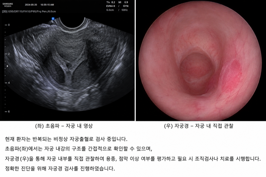
Most infertile women see their uterus only through transvaginal ultrasound. It is the starting point and the most important basic exam. It quickly and safely provides information on uterine size and shape, endometrial thickness, follicle growth, and fibroids. It is indispensable because it avoids radiation exposure and allows for repeat examinations.
However, an ultrasound is, after all, an image created using sound waves. Fertility doctors interpret grayscale cross-sectional images to judge the state of the uterus. While resolution has improved significantly, it cannot always distinguish minute surface changes inside the uterine cavity or tiny lesions only a few millimeters in size. Depending on the location, size, and state of the endometrium, abnormalities can often appear normal.
Hysteroscopy sees the uterus in an entirely different way. A thin endoscope, 3–5 mm in diameter, is inserted through the cervix, and the uterine cavity is gently expanded with saline. The moment the light at the tip of the camera turns on, the inside of the uterus—smaller than a palm—unfolds across the screen. The pink endometrium appears as a living tissue, and the base of the uterus where the embryo implants, along with the openings of the fallopian tubes, are clearly revealed. You can check the flow of blood vessels, the texture of the lining, and even the tiniest protrusions on an enlarged screen.
If an ultrasound is a satellite photo, a hysteroscope is like opening the front door and walking inside the house.
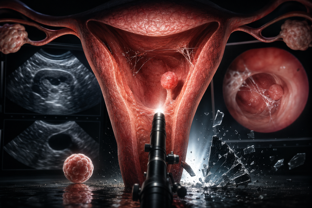
Therefore, lesions that appeared as mere shadows on an ultrasound can look completely different under a hysteroscope. A common example is an endometrial polyp. Most are benign, but if they are only 2–3 mm in size, they may not be distinguishable from the endometrium on an ultrasound. On the hysteroscopy screen, however, a tiny bump rising like a mushroom on the lining becomes clearly visible.
The same applies to uterine adhesions—as thin as a spider web. Adhesions from miscarriages or uterine surgeries might look normal on an ultrasound, but the hysteroscopy reveals the thread-like membranes connecting the uterine walls. If the adhesions are severe, they can narrow the uterine cavity and prevent the lining from growing sufficiently, potentially impacting the environment for implantation.
Beyond these, hysteroscopy is also crucial for diagnosing submucosal fibroids, congenital uterine septa, retained tissue after miscarriage, and suspected chronic endometritis.
Diagnosis and Treatment in One
Most importantly, hysteroscopy does not end with discovery. Polyps can be resected, adhesions can be lysed, and small septa can be corrected during the same procedure. Diagnosis and treatment become a single, unified process.
So why isn’t such an accurate test performed on every infertile patient? The answer is clear: while it is an excellent tool, it is not necessary for everyone. International guidelines from organizations like ESHRE and the NICE (UK) suggest there is insufficient evidence to show that routine hysteroscopy for women preparing for their first IVF—without abnormalities in basic tests—significantly increases birth rates.
While hysteroscopy is safely performed on an outpatient basis, it is an invasive procedure that can cause pain or discomfort, and rarely, complications like infection, bleeding, or uterine perforation can occur. The current standard is to select patients who will benefit most from the test rather than performing it on everyone.
The candidates for whom hysteroscopy is actively considered are clear: those with recurrent implantation failure or recurrent miscarriage, suspected polyps or submucosal fibroids on ultrasound, or suspected uterine adhesions/malformations. For these patients, hysteroscopy provides the decisive clue that can change the direction of treatment.
Conversely, one should be wary of excessive expectations. Many patients show no abnormalities during hysteroscopy, and removing a lesion does not guarantee pregnancy success. IVF success is determined by multiple factors, including maternal age, ovarian function, chromosomal state, sperm quality, and endometrial receptivity. Hysteroscopy is simply a way to more accurately piece together one specific part of that complex puzzle: the “uterine cavity.”
The uterus is the only space where an embryo must stay for nine months. The beginning of invisible life always happens in an invisible space. Looking directly into this space often provides the first clue explaining repeated failures.
※ This article was written based on the European Society of Human Reproduction and Embryology (ESHRE) guidelines, UK National Institute for Health and Care Excellence (NICE) recommendations, international medical papers, and consultations with domestic fertility specialists. It does not replace a specific individual’s diagnosis or treatment, and actual medical judgment must be made through consultation with a specialist.
※ Images: Created using generative AI (ChatGPT, OpenAI); depicts fictional individuals, not real people.